X-Ray Imaging
X-ray imaging is the oldest and most widely used form of medical imaging, and most people have one at some point in their lives.
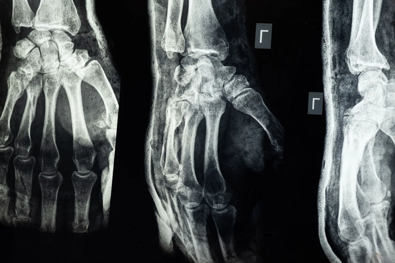
X-ray imaging is the oldest and most widely used form of medical imaging, and most people have one at some point in their lives. It is fast, painless, and especially good at showing bones. This guide explains what x-ray imaging is, why doctors order it, how the procedure works, how safe the radiation is, and why the report always belongs to a radiologist and your own doctor rather than to a web search.
The short version
- X-ray imaging, or radiography, uses a small dose of radiation to create pictures of the inside of the body.
- It is excellent for bones and the chest, and can also image the lungs, teeth, and, with contrast, the digestive tract.
- The radiation dose is low, and a doctor weighs the benefit against that small risk before ordering the test.
- A radiologist interprets the image and reports to your doctor, who explains what it means for you.
What x-ray imaging is
An x-ray is a form of high-energy radiation that can pass through the body. During imaging, a machine sends a controlled beam through the area of interest onto a digital detector on the other side. Dense tissues such as bone absorb more of the beam and appear white, while soft tissues and air absorb less and appear in shades of grey and black. The result is a two-dimensional picture that maps those differences in density. As MedlinePlus puts it, x-rays are a painless test that send a small amount of radiation through your body to create pictures of bones and organs.
X-ray imaging is one branch of a wider field. For the full map of scans that a hospital or clinic can offer, from ultrasound to MRI, see our overview of radiology.
Why a doctor orders an x-ray

X-ray imaging is often the first test used because it is quick, inexpensive, and widely available. According to MedlinePlus, common uses include finding broken bones, detecting pneumonia on a chest x-ray, and screening for breast cancer with a mammogram, and x-rays are also built into other tests such as CT scans. Typical reasons your doctor may request one include:
- Checking for fractures, dislocations, and joint problems after an injury.
- Looking at the lungs and heart when you have a cough, fever, or breathlessness.
- Investigating abdominal problems, such as a bowel blockage or swallowed object.
- Assessing teeth and jawbone in dentistry.
- Guiding and checking the position of implants, screws, or a healing bone.
Some studies focus on a specific area, such as a chest x-ray for suspected pneumonia or an abdominal film, and each has its own positioning and preparation.
How the procedure works, step by step
A plain x-ray is straightforward and usually takes only a few minutes. The exposure itself lasts a fraction of a second.
- You may be asked to remove clothing, jewellery, or metal objects from the area being imaged and put on a gown.
- The radiographer positions the body part between the x-ray machine and the detector plate.
- You hold still, and for a chest or abdominal film you may briefly hold your breath, while the image is captured.
- Several views from different angles are often taken so the radiologist can see the structure fully.
The NHS notes that you will not feel anything as the image is taken, and the whole process takes just minutes. Some specialised x-ray studies use a contrast agent, such as an injected dye or a barium drink, to make blood vessels or the digestive tract show up more clearly. The people who operate the equipment train along a defined path, described in our guide to the x-ray technologist role.
Types of x-ray imaging
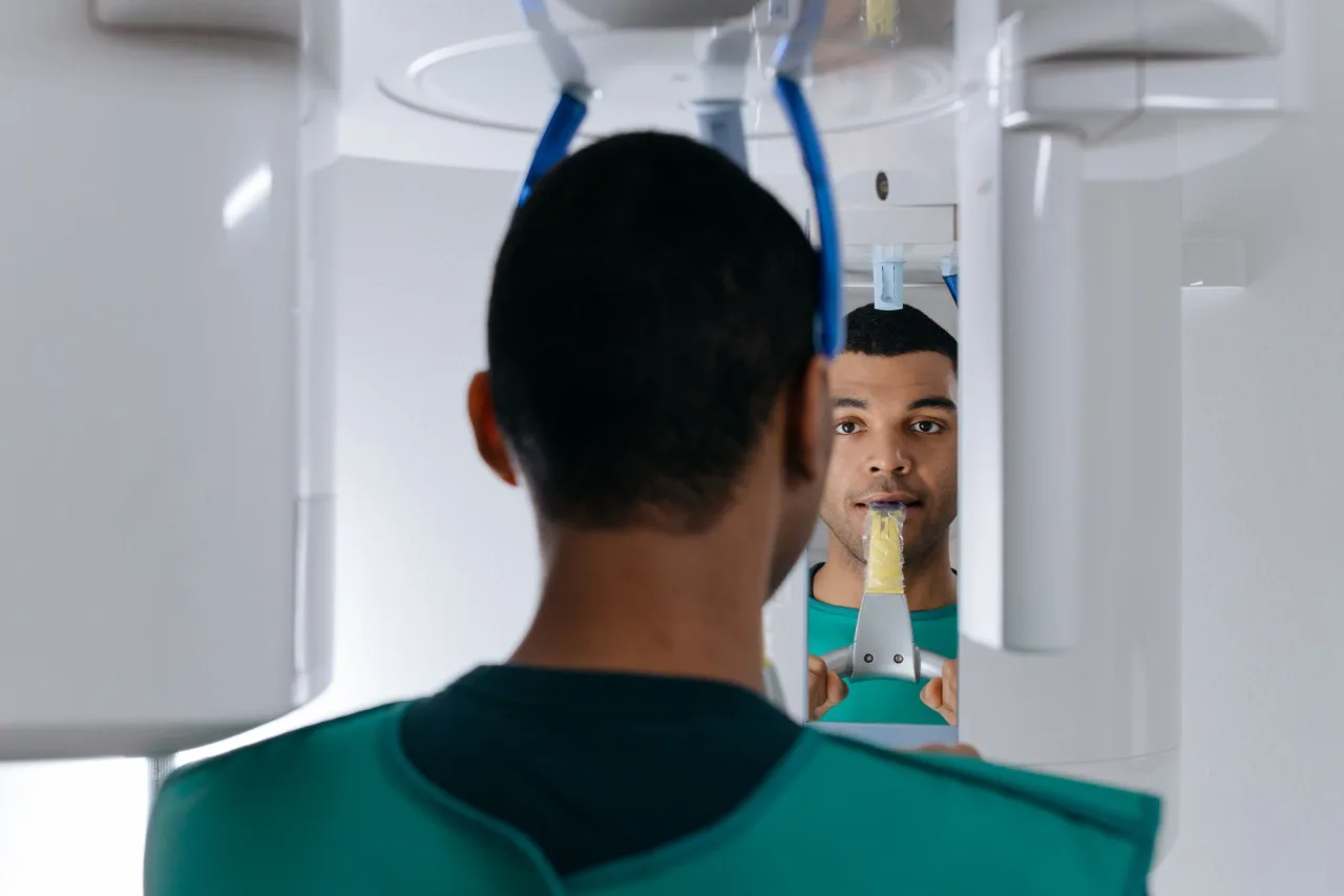
Not all x-ray imaging looks the same. A plain radiograph is the classic still picture of a bone or the chest. Fluoroscopy produces moving x-ray images in real time, used to watch the gut or guide a procedure. Mammography is a low-dose x-ray designed for breast tissue. Computed tomography, or CT, uses a rotating x-ray tube to build detailed cross-sectional slices, and our guide to the CT scan machine explains how that works. Knowing which type you are booked for helps you understand the preparation and the small differences in radiation dose.
Is the radiation safe?
The radiation dose from a single plain x-ray is low. MedlinePlus notes that a chest x-ray delivers roughly the same radiation as the natural background you receive from the environment over about 10 days. The NHS describes x-rays as generally safe and points out that a doctor assesses the risks before offering one, since there is a very small chance that radiation could contribute to cancer over many exposures.
Important: Tell your provider if you are pregnant or think you might be, because special precautions may apply. No radiation remains in your body after a plain x-ray, and the image itself is not radioactive. Facilities follow the principle of using the lowest dose that still answers the clinical question.
What your results mean
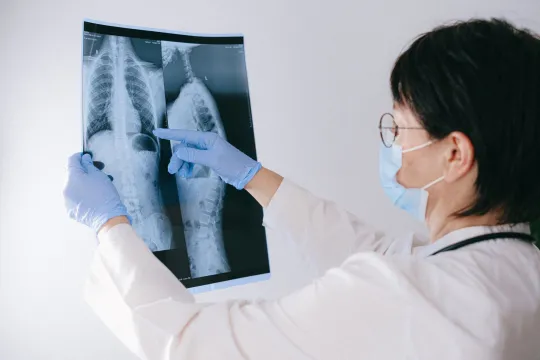
After the image is taken, a radiologist examines it and writes a report for the doctor who ordered the test. That doctor then explains the findings to you alongside your symptoms and history. A description such as a finding or an opacity is not automatically bad news, and many notes on a report turn out to be minor or long-standing.
X-ray imaging has genuine limits. It shows dense structures well but is far less sensitive to soft tissue detail than MRI or ultrasound, so a normal x-ray does not always rule out a problem. Reports are written for clinicians and often use cautious, technical language that can sound more alarming than the situation warrants. Try not to draw conclusions from an image or an online portal before your doctor has explained it.
This is general information, not medical advice. The same appearance on an x-ray can mean very different things depending on your age, symptoms, and history. Do not attempt to interpret your own image or report. Always discuss the findings with the doctor who ordered the scan or a specialist they refer you to.
Cost and where to have an x-ray
What an x-ray costs varies by country, by provider, by the body part imaged, and by whether you use insurance or pay directly, though it is usually one of the more affordable imaging tests. Confirm the price and your coverage with both the clinic and your insurer before booking. Choosing an accredited facility matters more than choosing the nearest, and our guide to finding an x-ray clinic near you explains what to look for. If your doctor decides you need more detail than a plain film can give, an MRI or CT scan may be the next step.
Frequently asked questions
What is x-ray imaging used for?
X-ray imaging is used to picture the inside of the body, most often to check for broken bones, examine the chest and lungs, screen for breast cancer with mammography, and look at the teeth. It is quick and widely available, which is why it is often the first imaging test a doctor orders.
Does an x-ray hurt?
No. An x-ray is painless, and you will not feel anything as the image is taken. You may need to hold an awkward position or hold your breath briefly so the picture is clear, but the exposure itself lasts only a fraction of a second.
How much radiation does an x-ray use?
The dose from a plain x-ray is low. A chest x-ray, for example, is roughly equal to about 10 days of natural background radiation. A doctor weighs the small risk against the benefit before ordering the test, and facilities use the lowest dose that still gives a useful image.
Can I have an x-ray if I am pregnant?
Tell your provider if you are or might be pregnant. An x-ray is sometimes still done with shielding and care, but your team will decide the safest approach and may consider a test that uses no radiation, such as ultrasound or MRI, instead. Always raise it before the scan.
Who explains my x-ray results?
A radiologist interprets the image and sends a report to the doctor who ordered it. That doctor then explains what the results mean for you. You should not rely on reading the image or report yourself, because interpretation depends on your full clinical picture.
More guides on MRI, CT & X-Ray
Have a question or a topic to suggest?
We read every message. Tell us what you would like to see next.
You may also like
MRI Tech
When people search for "MRI tech," they usually mean one of two things: the technology that makes a magnetic resonance imaging scan possible, or the technologist who runs the machine.

MRI Testing
MRI testing can feel mysterious the first time: a referral appears, an appointment gets booked, and you are told to lie still inside a loud tunnel.
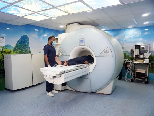
Stand Up MRI
A stand up MRI, sometimes called an upright, positional or weight-bearing MRI, lets you be scanned sitting, standing or bending rather than only lying flat.