MRI Tech
When people search for "MRI tech," they usually mean one of two things: the technology that makes a magnetic resonance imaging scan possible
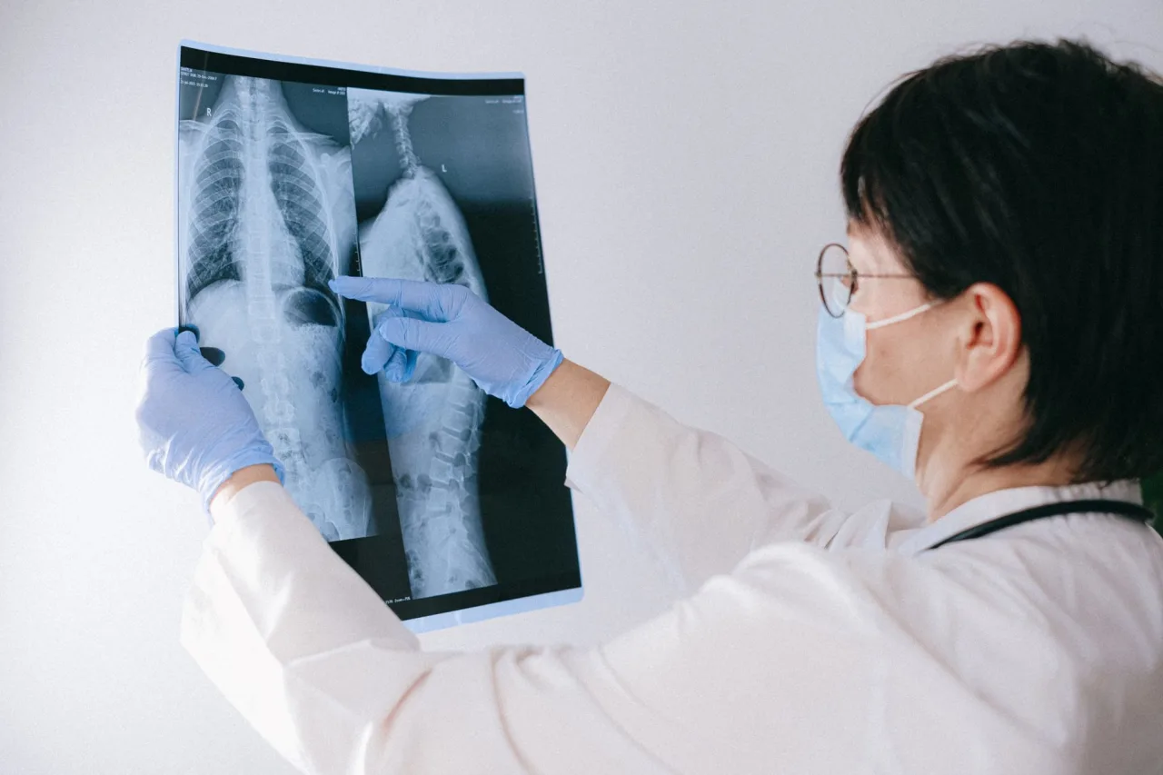
When people search for "MRI tech," they usually mean one of two things: the technology that makes a magnetic resonance imaging scan possible, or the technologist who runs the machine. This guide covers the technology in plain language, explains how the scanner turns magnets and radio waves into detailed pictures, and points you to the right places to prepare, compare costs and choose a clinic.
The short version
- MRI technology uses a strong magnet, radio waves and a computer to build images. It does not use ionizing radiation like X-rays or CT.
- Magnet strength is measured in tesla (T). Most clinical scanners are 1.5T or 3T, and higher field strength can mean sharper images for some studies.
- Scanners come in several shapes: closed (traditional bore), wide-bore and open designs that help with claustrophobia or larger body sizes.
- A radiologist, not the machine or the technologist, interprets the images and sends a report to the doctor who ordered your scan.
What "MRI tech" actually refers to
Magnetic resonance imaging is a way of looking inside the body without surgery and without radiation. According to MedlinePlus, an MRI "uses magnets, radio waves, and a computer to create detailed pictures of the inside of your body," and it "does not use x-rays or other radiation," which is why it can be repeated more freely than some other tests. The "tech" in MRI tech can point at the equipment itself or at the trained MRI technologist who positions you, runs the sequences and keeps you safe around the magnet.
This article focuses on the equipment and how it works. If you want the full patient walk-through of being referred, scanned and getting a report, read our companion guide to MRI testing, procedure and results, and for the core medical overview see what an MRI scan is.
How the technology creates an image
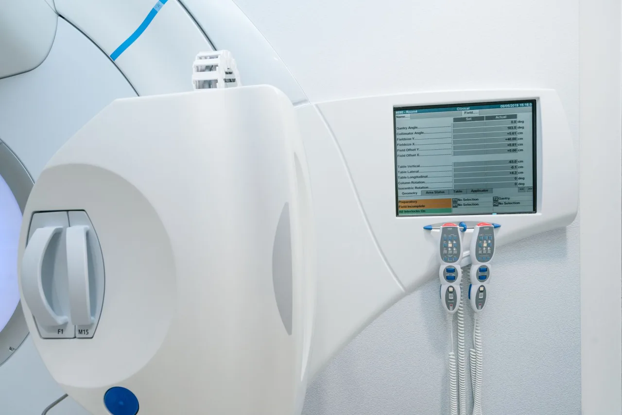
The human body is mostly water, and water contains hydrogen. RadiologyInfo, the patient resource from the American College of Radiology and the Radiological Society of North America, explains that an MRI unit uses "a powerful magnetic field, radiofrequency pulses, and a computer to produce detailed pictures." In simple terms, the strong magnet lines up hydrogen atoms in your tissues, a radio wave pulse nudges them out of alignment, and as they settle back they release small signals. Antennas called coils detect those signals, and the computer reconstructs them into cross-sectional images.
Because different tissues release signals at slightly different rates, MRI is very good at showing soft tissue such as the brain, spinal cord, muscles, ligaments and organs. It is often the test of choice when doctors need to tell healthy tissue from a problem area that an X-ray would miss.
Magnet strength: what 1.5T and 3T mean
Magnet strength is measured in units called tesla. Most hospital and outpatient scanners run at 1.5 tesla or 3 tesla. A 3T magnet is roughly twice as strong as a 1.5T magnet and can produce higher-resolution images or shorter scans for certain studies, such as detailed brain, joint or vascular imaging. A 1.5T scanner remains an excellent, widely used workhorse and is sometimes preferred for patients with certain implants.
Stronger is not automatically better for every case. The right field strength depends on the body part, the clinical question and any implanted devices you carry. The radiologist and technologist choose the scanner and settings for your specific study.
Types of MRI machines
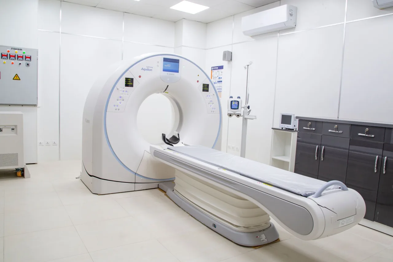
- Closed or traditional bore: a cylinder with a relatively narrow tunnel. It often gives the strongest, most uniform magnetic field and the best image quality.
- Wide-bore: a larger opening that keeps high field strength while giving more room, which helps with claustrophobia and larger body habitus.
- Open MRI: designs with open sides for anxious or claustrophobic patients. RadiologyInfo notes that open units with larger diameters can accommodate claustrophobic patients, though some exam types still need a traditional design.
If enclosed spaces worry you, it can be worth searching specifically for an open MRI facility near you and asking whether it can perform the study your doctor ordered.
A note on safety around the magnet
The magnet is always on, even between scans. That is why metal is taken so seriously. The NHS advises removing jewellery, some clothing with metal and hearing aids, and staff will ask about pacemakers, aneurysm clips, cochlear implants and surgical hardware before you enter the room. Always tell the technologist about every implant, and mention if you might be pregnant.
What the machine is used to look at
MRI technology is versatile. RadiologyInfo lists uses including evaluating the abdomen and pelvis, blood vessels and lymph nodes, and helping diagnose tumours, liver disease and inflammatory conditions. MedlinePlus adds common musculoskeletal uses such as the knee, shoulder and other joints, plus the brain, spinal cord and nerves. Because there is no ionizing radiation, MRI is also used carefully in situations where repeat imaging is needed.
When doctors need to compare tools, they may weigh MRI against a CT study. Our overview of CT and MRI together explains where each one shines.
The role of contrast material
Some MRI studies use a contrast agent, most often based on gadolinium, given through a vein to highlight blood vessels, inflammation or certain tumours. RadiologyInfo notes gadolinium is generally well tolerated and is an option for people with iodine allergy, but that a rare condition called nephrogenic systemic fibrosis has been linked to its use in patients with serious kidney disease. Your care team screens kidney function when contrast is planned. Whether you need contrast is a medical decision made by the radiologist and your doctor.
What happens during a scan
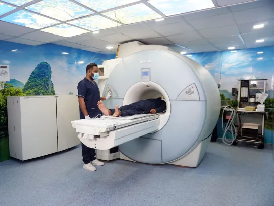
You lie on a table that slides into the bore. A coil may be placed over the body part being studied. The scan is loud, so you are given ear protection, and you must hold very still because motion blurs the images. A typical study runs from about 15 to 60 minutes depending on how many sequences are needed. The technologist watches and talks to you from the control room, and you usually hold a call button.
Understanding your results
The scanner does not diagnose anything. It produces images that a radiologist reviews, then writes a report for the clinician who ordered the test. That report describes what the images show and often suggests next steps. Do not try to read the pictures yourself or interpret a preliminary impression as a final answer. The meaning of any finding depends on your symptoms, history and other tests, which is why the conversation with your own doctor matters most.
If cost is on your mind, imaging prices vary widely, so it helps to review typical MRI prices with and without insurance and to call the MRI center directly for a quote before you book.
This is general information, not medical advice. Everything here is educational. It cannot tell you whether you need an MRI, which scanner is right for you, or what your specific images mean. Always talk with your referring clinician or a radiologist about your own situation.
Frequently asked questions
Does an MRI use radiation?
No. MRI uses a strong magnet and radio waves, not ionizing radiation. MedlinePlus states that MRI does not use x-rays or other radiation, which is one reason it may be chosen when repeat imaging is needed. This is different from X-ray and CT, which do use radiation.
What is the difference between a 1.5T and a 3T MRI?
The number refers to magnet strength in tesla. A 3T scanner has a stronger field than a 1.5T scanner and can give higher-resolution images or faster scans for some studies. Neither is best for every case, and the right choice depends on the body part, the clinical question and any implants you have. Your radiologist decides.
Is an MRI technologist the same as a radiologist?
No. The MRI technologist is the trained professional who positions you, operates the scanner and keeps you safe. A radiologist is a medical doctor who interprets the images and writes the report. Both are part of your imaging team, but only the radiologist provides the diagnostic reading.
Can I have an MRI if I have metal in my body?
It depends on the type of metal or device. Some implants, such as certain pacemakers, aneurysm clips and cochlear implants, can be unsafe or affected by the magnet. Always disclose every implant and surgery, and let the staff decide whether the scan is safe for you.
Why is the MRI machine so loud?
The knocking and buzzing come from the scanner's coils switching rapidly during imaging. It is normal. Clinics provide earplugs or headphones, and you can usually listen to music or speak to the technologist between sequences.
More guides on MRI, CT & X-Ray
Have a question or a topic to suggest?
We read every message. Tell us what you would like to see next.
You may also like

MRI Testing
MRI testing can feel mysterious the first time: a referral appears, an appointment gets booked, and you are told to lie still inside a loud tunnel.
Stand Up MRI
A stand up MRI, sometimes called an upright, positional or weight-bearing MRI, lets you be scanned sitting, standing or bending rather than only lying flat.

X-Ray Scan
An X-ray scan is one of the fastest and most common medical imaging tests, often done in minutes to check bones, the chest and more. This guide explains what an X-ray exam involves, why doctors order it