Chest X-Ray of Pneumonia
If you have a fever, a cough, and breathlessness, one of the first tests a doctor may order is a chest x-ray.
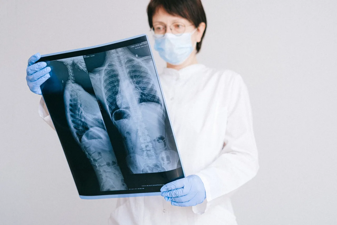
If you have a fever, a cough, and breathlessness, one of the first tests a doctor may order is a chest x-ray. It is a fast, painless way to look at the lungs and check for signs of pneumonia. This guide explains why the scan is used for a suspected chest infection, how it is done, what a radiologist looks for, and why only your doctor can tell you what your own images mean.
The short version
- A chest x-ray uses a very small dose of radiation to picture the lungs, heart, and chest wall.
- Doctors use it to help confirm pneumonia and to see how much of the lung is affected.
- A normal x-ray does not always rule out infection, and an abnormal shadow is not always pneumonia.
- A radiologist reads the image and reports to your doctor, who explains what it means alongside your symptoms and tests.
What a chest x-ray is
A chest x-ray, also called chest radiography, is the most common imaging test of the chest. A machine sends a small, controlled beam of x-rays through your body onto a detector, and the different tissues absorb the beam by different amounts. Bone absorbs a lot and appears white, air-filled lungs absorb little and appear dark, and fluid or infection shows up as paler, cloudy areas. According to RadiologyInfo.org, the test evaluates the lungs, heart, and chest wall and helps diagnose symptoms such as breathing difficulty, a persistent cough, chest pain, and fever.
Pneumonia is an infection that inflames the tiny air sacs in one or both lungs, and they can fill with fluid or pus. On an x-ray, that fluid replaces the normal dark air pattern with a whiter, hazy patch that a radiologist may describe as a consolidation or an infiltrate. For a broader view of how this test fits among other radiology studies, see our overview of x-ray imaging.
Why a doctor orders a chest x-ray for suspected pneumonia
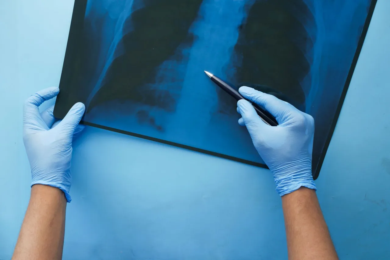
Pneumonia can be difficult to tell apart from a cold, flu, or bronchitis by symptoms alone. As MedlinePlus explains, a provider usually starts with your medical history, listens to your lungs with a stethoscope, and then orders tests, and a chest x-ray is a primary tool to confirm the diagnosis. Common reasons your doctor may request one include:
- Confirming whether a chest infection has reached the lungs rather than staying in the airways.
- Judging how much of the lung is involved and whether one or both lungs are affected.
- Looking for complications such as fluid collecting around the lung, known as a pleural effusion.
- Ruling out other causes of the same symptoms, such as heart failure or, rarely, a lung mass.
- Checking whether the infection is clearing after treatment, if your recovery is slow.
In more serious or hospital cases, MedlinePlus notes that a CT scan may be added to see how much of the lung is affected. Our guide to the CT scan machine explains how that more detailed test works.
How the scan works, step by step
A chest x-ray is quick and needs little preparation. The whole appointment usually takes about 15 minutes, and the exposure itself lasts a fraction of a second.
- You remove clothing and jewellery from the upper body and put on a gown, since zips and metal show up on the image.
- You stand with your chest against the flat detector plate, with the x-ray machine behind you.
- The radiographer usually takes two views, one from back to front and one from the side, as RadiologyInfo.org describes.
- You take a deep breath and hold still for a moment while the image is captured, because full lungs give the clearest picture.
You will not feel anything during the exposure. If you cannot stand, the image can be taken sitting up or lying down using a portable machine, which is common on hospital wards. The technologists who run these machines follow a defined training path, described in our look at the x-ray technologist role.
Is the radiation safe?
The dose from a single chest x-ray is very small. MedlinePlus notes that the radiation from a chest x-ray is roughly the same as the natural background radiation you receive from the environment over about 10 days. The NHS describes x-rays as generally safe, while acknowledging a very small risk that radiation could contribute to cancer over many exposures, which is why a doctor weighs the benefit of each scan first.
Important: Always tell the clinic if you are pregnant or think you might be. A chest x-ray is often still possible with shielding, but your care team will decide the safest approach for you. No radiation stays in your body after the exposure ends.
What the results can and cannot show
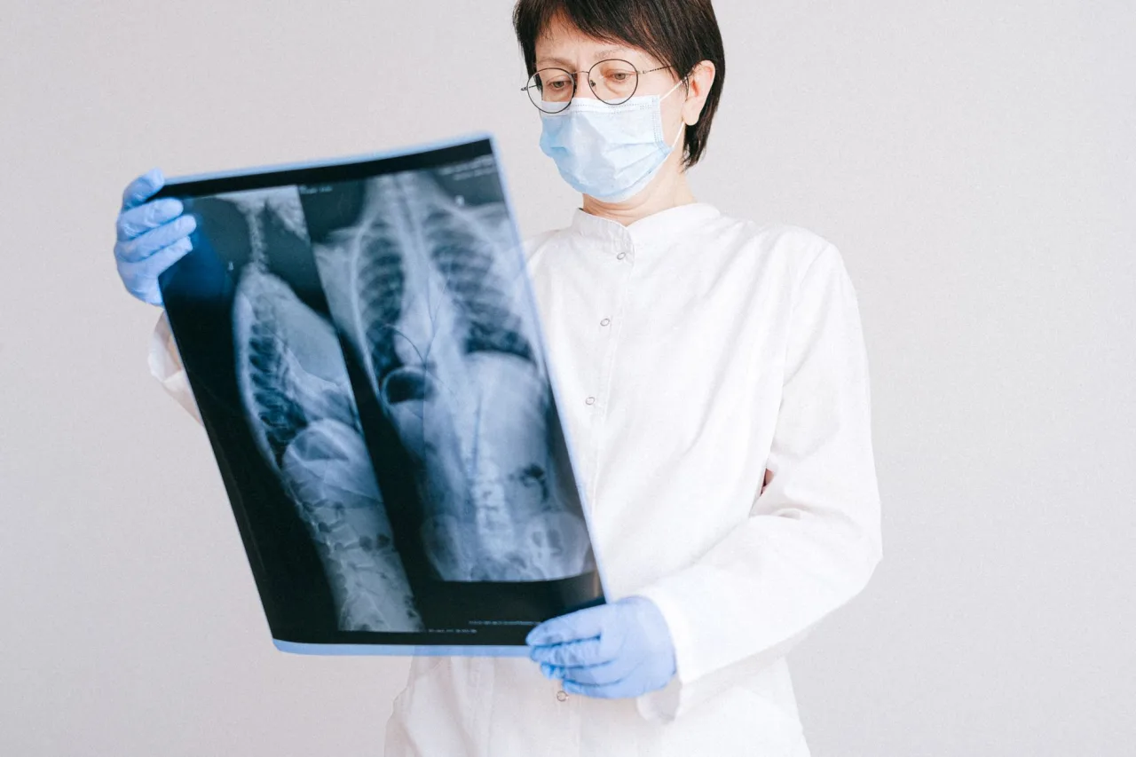
After the scan, a radiologist studies the image and writes a report for the doctor who ordered it. When pneumonia is present, they often see a cloudy white area in part of the lung. They also note whether one area or several are involved, whether there is fluid around the lung, and whether the heart and other structures look normal.
An x-ray has real limits, though. RadiologyInfo.org points out that a chest x-ray cannot detect every problem, and small cancers or blood clots in the lung may not appear. Early pneumonia can also look normal on a first x-ray, and changes sometimes lag a day or two behind symptoms. Equally, a shadow on the film is not automatically pneumonia, because scarring, old infection, or other conditions can look similar. This is exactly why the image is read alongside your symptoms, examination, and blood tests rather than on its own.
This is general information, not medical advice. A chest x-ray showing a shadow does not tell you, by itself, what is wrong or how serious it is. Do not try to interpret your own image or an online report. Always discuss the findings with the doctor who ordered the scan, and seek urgent care if you are severely breathless or your lips turn blue.
After the x-ray: treatment and follow-up
If your doctor confirms pneumonia, treatment depends on the cause and how unwell you are. Bacterial pneumonia is usually treated with antibiotics, while rest, fluids, and time help the body recover. Many people are treated at home, but some need hospital care, particularly older adults, young children, and those with other health conditions.
Your doctor may or may not order a repeat x-ray. A follow-up film is sometimes done a few weeks later, especially in older adults or smokers, to confirm the lung has cleared and that nothing else was hidden behind the infection. If your symptoms are not improving as expected, that follow-up becomes more important. Never stop or change prescribed treatment based on how you feel alone without speaking to your clinician first.
Cost and where to have a chest x-ray
What a chest x-ray costs varies widely by country, provider, and whether you use insurance or pay directly, and it is usually one of the cheaper imaging tests. Always confirm the price and your coverage with both the clinic and your insurer before booking. Choosing an accredited facility matters more than choosing the nearest one, and our guide to finding an x-ray clinic near you explains what to check. For a wider view of imaging tests and how they relate, see our guide to radiology.
Frequently asked questions
Can a chest x-ray always detect pneumonia?
Not always. A chest x-ray is a primary test for pneumonia, but very early infection can look normal, and changes sometimes appear a day or two after symptoms start. Your doctor reads the image alongside your examination and blood tests, and may repeat the x-ray or order a CT scan if the picture is unclear.
What does pneumonia look like on a chest x-ray?
Pneumonia usually shows up as a cloudy, whiter area where healthy lung would normally look dark, because the air sacs fill with fluid or pus. A radiologist may call this a consolidation or an infiltrate. Only a trained clinician can confirm what any shadow means in your case.
How long does a chest x-ray take?
The appointment usually takes about 15 minutes, and the exposure itself lasts a fraction of a second. Most of the time is spent changing into a gown and positioning you correctly against the detector plate for a clear picture.
Is the radiation from a chest x-ray dangerous?
The dose is very small, roughly equal to about 10 days of natural background radiation. It is considered low risk when the scan is medically justified. Tell the clinic if you are or might be pregnant so they can take extra care, and discuss any concerns with your doctor.
Who tells me the results of my chest x-ray?
A radiologist interprets the image and sends a report to the doctor who ordered it. That doctor then explains what it means for you in the context of your symptoms and other tests. You should not rely on reading the image or report yourself.
More guides on MRI, CT & X-Ray
Have a question or a topic to suggest?
We read every message. Tell us what you would like to see next.
You may also like

Sonography Machine Price: Real Prices
Search "sonography machine price" and you land on two very different questions: what does it cost a clinic to buy the scanner, and what will a patient pay for the scan it produces?
Ultrasound Device Price: Real Prices
"Ultrasound device" covers a lot of ground, from a wireless probe that plugs into a phone to a room-sized diagnostic console. Because the category is so broad, so is the price.
Ultrasound Scan Machine Cost: Real Prices
"Ultrasound scan machine cost" hides two questions inside one phrase: what a clinic pays to own and run the machine, and what you pay for the scan it produces.