MRI Abdomen
An MRI of the abdomen produces exceptionally detailed pictures of the organs in the belly
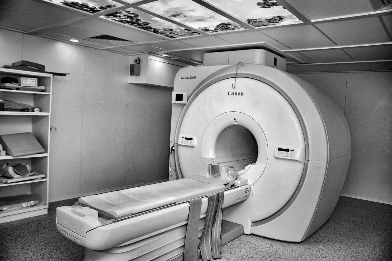
An MRI of the abdomen produces exceptionally detailed pictures of the organs in the belly, using a strong magnet and radio waves rather than any radiation. It is often used when a doctor needs a closer look at the liver, kidneys, pancreas, or other structures. This guide explains why an abdominal MRI is ordered, how the scan works, how to prepare, what contrast does, and why only a radiologist and your doctor can interpret the results.
The short version
- An abdominal MRI uses a strong magnet and radio waves, not radiation, to image organs such as the liver, kidneys, and pancreas.
- The scan is painless but longer and noisier than a CT, usually taking around 30 to 60 minutes.
- A contrast dye called gadolinium is sometimes injected to make organs and vessels clearer.
- A radiologist reads the images and reports to your doctor, who explains what they mean for you.
What an abdominal MRI is
MRI stands for magnetic resonance imaging. The scanner is a large cylindrical magnet with a bed that slides into the centre. Instead of x-rays, it uses a powerful magnetic field and pulses of radio waves to make the water molecules in your body give off signals, which a computer turns into highly detailed cross-sectional images. As RadiologyInfo.org explains, body MRI evaluates abdominal and pelvic organs including the liver, kidneys, and pancreas, as well as blood vessels and lymph nodes, and it uses magnetic fields and radio waves rather than radiation.
Because it excels at soft tissue detail, an abdominal MRI can show things that other tests may miss or leave unclear. For a wider view of how it compares with other scans, see our overview of radiology, and for the equipment behind the faster alternative, our guide to the CT scan machine.
Why a doctor orders an abdominal MRI
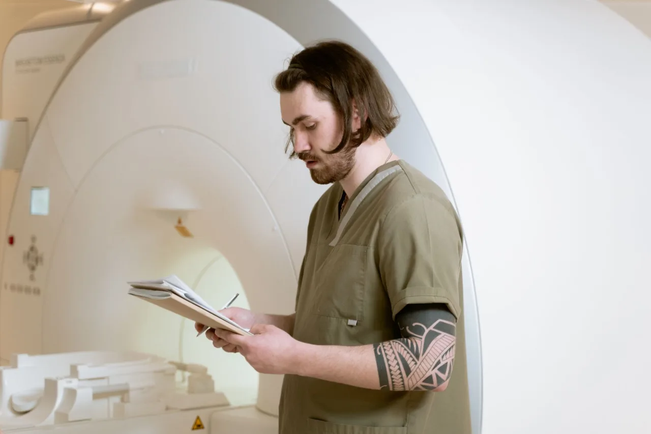
An abdominal MRI is often used to look more closely at something found on an earlier ultrasound or CT, or to answer a specific question about the soft organs. Common reasons include:
- Characterising a liver lesion or assessing liver disease such as fatty liver or cirrhosis.
- Examining the bile ducts and pancreas, sometimes with a specialised sequence called MRCP.
- Assessing the kidneys and adrenal glands, and staging some cancers.
- Investigating bowel inflammation, such as in Crohn's disease.
- Looking at blood vessels and lymph nodes in the abdomen.
Because it uses no ionising radiation, MRI is sometimes preferred for younger patients or when repeated imaging is likely. If the area of interest is lower down, your doctor may instead request an MRI of the pelvis.
How to prepare
Preparation depends on the exact scan, and the clinic will give you specific instructions. RadiologyInfo.org advises removing all jewellery and metallic items, wearing loose comfortable clothing, following any food and drink guidance for your particular exam, and disclosing all medical implants and any metal in your body.
Important: The MRI magnet is always on. Tell the clinic in advance if you have a pacemaker, cochlear implant, aneurysm clip, or any other implant or metal fragment, because some are not compatible with the scanner. The NHS notes that you must declare surgical clips, a pacemaker, dental work, and similar items before an MRI. Your care team will confirm what is safe for you.
What happens during the scan, step by step

An abdominal MRI is painless but takes longer than most scans. RadiologyInfo.org puts a typical body MRI at around 30 to 60 minutes.
- You change into a gown and remove all metal, then lie on the padded bed, usually on your back.
- If contrast is needed, a small cannula is placed in a vein for a gadolinium injection during the scan.
- You are given earplugs or headphones, because the machine makes loud knocking and clicking sounds during imaging.
- The bed slides into the tunnel, and you hold still, sometimes holding your breath briefly, while each sequence runs.
A radiographer watches and speaks to you from the next room, as the NHS describes, and you are given a buzzer to press if you need to stop. The tunnel is more enclosed than a CT ring, so tell the clinic beforehand if you are claustrophobic, as options such as an open scanner or mild sedation may be available.
Contrast dye: what it does
Some abdominal MRI scans use an injection of contrast material to make organs, blood vessels, or areas of inflammation stand out more clearly. RadiologyInfo.org notes that MRI contrast is usually a gadolinium-based agent, which is generally safer for people with allergies than the iodine-based dye used in CT. Your kidney function may be checked with a blood test first, since the kidneys clear the dye.
Serious reactions to gadolinium are uncommon, but tell the clinic if you have had a reaction before, have kidney problems, or are pregnant or breastfeeding. Your radiologist will decide whether contrast is needed and which agent is appropriate for you.
What your results mean
After the scan, a radiologist studies the images and writes a report for the doctor who ordered the test. That doctor then explains the findings to you in the context of your symptoms, examination, and any other tests. A result described as a finding is not automatically serious, and many incidental findings turn out to be harmless.
How quickly you get results depends on the setting, from a same-day report in an urgent case to a week or more for a routine outpatient scan. It is normal to feel anxious in the gap, but try not to interpret the images or an online portal yourself. MRI reports are written for clinicians and use cautious, technical language that can sound more alarming than the situation warrants.
This is general information, not medical advice. Abdominal MRI images are complex, and the same appearance can mean very different things depending on your situation. Do not attempt to interpret your own scan or report. Always discuss the findings with the doctor who ordered the scan or a specialist they refer you to.
Cost and where to have an abdominal MRI
What an abdominal MRI costs varies widely by country, by provider, by whether contrast is used, and by whether you use insurance or pay directly, and MRI is generally one of the more expensive imaging tests. Confirm the price and your coverage with both the clinic and your insurer before booking, and see our guide to MRI prices for realistic ranges. Choosing an accredited clinic matters more than choosing the nearest, and our guide to finding an MRI centre near you explains what to check.
Frequently asked questions
Does an abdominal MRI use radiation?
No. An MRI uses a strong magnetic field and radio waves, not ionising radiation, to create its images. This is one reason it may be chosen over CT for some patients, particularly when repeated imaging is likely or radiation is a concern. Discuss the best option with your doctor.
How long does an abdominal MRI take?
A body MRI typically takes around 30 to 60 minutes, longer than a CT scan. The exact time depends on how many sequences are needed and whether contrast is used. You need to lie still throughout, and the machine is noisy, so you will be given earplugs or headphones.
Do I need contrast dye for an abdominal MRI?
It depends on the reason for your scan. Contrast, usually a gadolinium-based dye, helps show organs, blood vessels, and inflammation more clearly, but not every scan needs it. Your clinic will tell you in advance and may check your kidney function first. Always mention past reactions and any kidney problems.
Is an abdominal MRI safe if I have metal in my body?
The MRI magnet is always on, so some implants and metal fragments are not compatible with the scanner. Tell the clinic beforehand about any pacemaker, cochlear implant, aneurysm clip, surgical clips, or metal in your body. Your care team will confirm whether an MRI is safe for you or whether another test is better.
Who explains my abdominal MRI results?
A radiologist interprets the images and sends a report to the doctor who ordered the scan. That doctor then explains what the results mean for you. You should not rely on reading the images or report yourself, because interpretation depends on your full clinical picture.
More guides on MRI, CT & X-Ray
Have a question or a topic to suggest?
We read every message. Tell us what you would like to see next.
You may also like
Fluoroscopy
Fluoroscopy is a form of medical imaging that turns X-rays into a live, moving picture of the inside of your body. Instead of a single still photo, your care team watches organs, joints
Hip X-Ray
A hip X-ray is a quick, low-dose imaging test that shows the bones of the hip joint and upper thigh. Doctors use it to look for fractures, arthritis
Cect Scan
CECT stands for contrast-enhanced computed tomography, a CT scan performed with a contrast dye to make organs, blood vessels and abnormal areas stand out more clearly.