X-Ray Tech: Uses, Procedure & Results
Why X-rays are ordered, how the test works, whether it is safe, and who performs and interprets your images.

"X-ray tech" is a phrase people use to mean two related things: the X-ray technology itself, the everyday imaging test that looks inside the body, and the radiologic technologist, the trained professional who actually performs it. This guide covers both. It explains why an X-ray is ordered, how the procedure works, whether it is safe, and how to think about your results, always with the reminder that only your own clinician can interpret your images for you.
The short version
- An X-ray is a fast, painless test that uses a small amount of ionizing radiation to image bones and some soft tissues.
- It is performed by a radiologic technologist and interpreted by a radiologist, a doctor who specialises in reading images.
- The radiation dose from a typical X-ray is low, but always tell staff if you are or might be pregnant.
- Your report describes what the images show; what it means for you is a conversation for your referring doctor.
What "X-ray tech" actually means
In casual search terms, "x ray tech" and "xray tech" get used for the machine, the image, and the person operating it. It helps to separate them. The technology is a form of medical imaging; the test is what your doctor orders; and the technologist, sometimes called a radiographer, is the credentialed professional who positions you and captures the images. According to MedlinePlus, an X-ray is a painless imaging test that sends a small amount of radiation through the body to create pictures of bones and organs, which a radiologist then reviews and reports to your provider.
If you are researching the career rather than the test, the short version is that in the United States the technologist role is credentialed by the American Registry of Radiologic Technologists (ARRT), which certifies professionals across radiography and related imaging disciplines after they meet education, examination and ethics requirements.
Why an X-ray is ordered
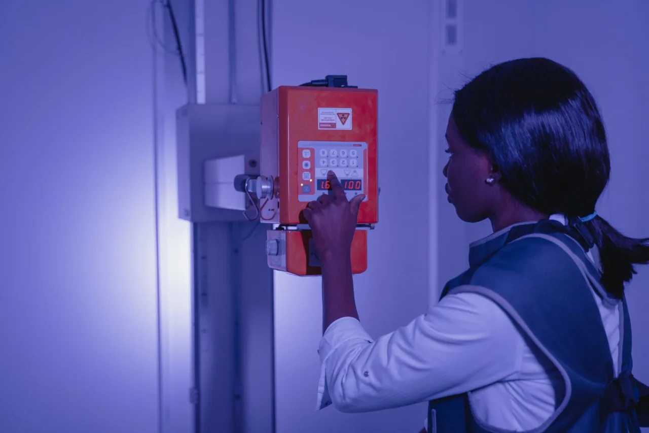
Doctors order X-rays because they are quick, widely available and very good at showing dense structures. Common reasons include:
- Bones and joints: checking for fractures, dislocations, arthritis or bone infections.
- Chest: looking at the lungs and heart, for example when pneumonia is suspected. Our guide to chest radiography covers this in more depth.
- Abdomen: assessing bowel gas patterns or locating swallowed objects.
- Screening and guidance: mammography is a specialised X-ray of the breast, and X-rays also help place or check medical devices. See our overview of the mammogram procedure for that context.
When an X-ray does not show enough detail, a doctor may follow up with a more detailed cross-sectional test such as a CT scan. Our explainer on what a CT scan involves describes when that step makes sense.
How an X-ray works
X-rays are a form of energy, like light and radio waves, but with enough energy to pass through the body. As they travel through you, dense tissues such as bone absorb more of the beam and appear white on the image, while air-filled areas such as the lungs appear dark, and soft tissues sit in shades of grey. A detector on the far side captures the pattern that gets through, and that pattern becomes the picture your radiologist reads.
Types of X-ray exam
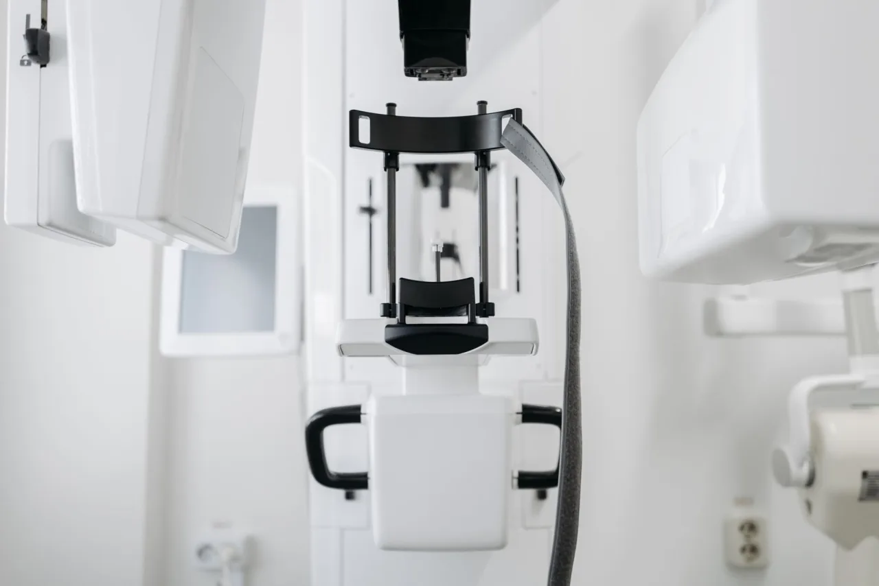
"X-ray" covers a family of related tests, not just the single still image most people picture. Depending on what your doctor needs to see, you might have:
- Plain radiography: the standard still image of bones, chest or abdomen.
- Fluoroscopy: a moving, real-time X-ray used to watch a structure in action or guide a procedure.
- Contrast studies: exams where a contrast agent, swallowed or injected, highlights specific structures such as the digestive tract or blood vessels.
- Mammography: a low-dose X-ray specialised for breast tissue.
- CT: a computer combines many X-ray images into detailed cross-sections, a step up in detail described in our CT scan guide.
Because these vary in preparation and dose, the instructions you are given depend on the exact exam, which is why it helps to read your appointment letter and ask the clinic if anything is unclear.
The procedure: what to expect
A plain X-ray is usually quick and straightforward. Most exams take only a few minutes for the imaging itself.
- Preparation: you may be asked to remove jewellery, glasses or clothing over the area being imaged and to wear a gown. Most plain X-rays need no fasting or special preparation.
- Positioning: the technologist positions the body part between the machine and the detector, and may use supports to hold you still.
- The exposure: you hold still, and sometimes hold your breath for a moment, while the image is taken. It is painless and you feel nothing.
- Multiple views: several images from different angles are often needed for a full picture.
Important
Always tell the technologist if you are pregnant or think you might be. As MedlinePlus notes, you should tell your provider about a possible pregnancy before an X-ray so they can weigh the benefits against any risk and adjust the approach if needed.
Is an X-ray safe?
X-rays use ionizing radiation, but the dose from a typical exam is small. RadiologyInfo.org notes that a single chest X-ray delivers roughly 0.1 millisieverts, about the same as the natural background radiation most people receive over about 10 days. Imaging teams also follow the ALARA principle, "as low as reasonably achievable", to keep exposure to a minimum. For most people the diagnostic benefit of a properly ordered X-ray clearly outweighs the small radiation risk, but that judgement belongs to your doctor, especially during pregnancy or for repeated imaging.
Who performs and reads your X-ray
Two different professionals are involved. The radiologic technologist runs the equipment, positions you and produces high-quality images while protecting you from unnecessary exposure. The radiologist, a medical doctor with specialist training, then interprets those images and writes the report that goes to your referring clinician. Many outpatient X-rays are done at radiology practices and imaging centres, a setup we describe in our guide to radiology associates and imaging groups.
If you are researching "x ray tech" as a career rather than as a patient, the technologist route in the United States generally runs through an accredited radiography programme, often an associate degree, followed by certification. The ARRT sets the credentialing standards and, as it describes, awards credentials only after candidates meet education, examination and ethics requirements, then requires ongoing continuing education to keep them current. Many technologists later add postprimary credentials in areas such as CT, MRI or mammography to broaden their scope. State licensing rules vary, so anyone planning this path should check the requirements where they intend to work.
What your results mean
After the exam, the radiologist reviews the images and produces a written report describing what they see, such as a fracture, a clear chest, or an area needing further tests. That report goes to the doctor who ordered the X-ray, who puts it together with your symptoms and history. It is genuinely important not to try to self-diagnose from an image or a report you find hard to read, because findings need clinical context to make sense.
A note on medical advice
This article is general educational information, not medical advice, and it cannot tell you what your own X-ray shows. For anything specific to you, speak with the doctor who ordered the test or the radiologist who reported it.
When you are ready to book, our guide to how to find and choose an X-ray clinic explains what to look for in an accredited, well-run facility.
Frequently asked questions
Does an X-ray hurt?
No. A plain X-ray is painless and you feel nothing during the exposure. The only mild discomfort some people notice is holding a position or staying still for a moment while the image is taken.
How much radiation is in an X-ray?
The dose is small. RadiologyInfo.org notes that a single chest X-ray delivers roughly 0.1 millisieverts, about the same as the natural background radiation most people receive over about 10 days. Imaging teams follow the ALARA principle to keep exposure as low as reasonably achievable.
Can I have an X-ray if I am pregnant?
Always tell the technologist if you are pregnant or think you might be. X-rays are sometimes still done in pregnancy when necessary, but your doctor will weigh the benefits against any risk and may adjust or postpone the test, so this is a decision to make with your provider.
Who reads my X-ray and how do I get the results?
A radiologist, a doctor who specialises in interpreting medical images, reviews your X-ray and writes a report. That report goes to the clinician who ordered the test, who then discusses the findings and any next steps with you.
What is the difference between an X-ray tech and a radiologist?
The radiologic technologist is the trained professional who operates the equipment, positions you and captures the images. The radiologist is a medical doctor who interprets those images and writes the diagnostic report. In the United States, technologists are credentialed by the ARRT.
More guides on MRI, CT & X-Ray
Have a question or a topic to suggest?
We read every message. Tell us what you would like to see next.
You may also like

X-Ray Near Me: How to Choose
An honest guide to finding an x-ray near you: how to choose an accredited clinic where a radiologist reports your images, what an x-ray costs, the small radiation dose, and the questions to ask first.

CT Scan: Uses, Procedure and Results
A CT scan, or CAT scan, uses x-rays and a computer to create detailed cross-sectional images of the body. Here is why doctors order one, how the procedure works, what contrast dye does, and how the radiation compares.
Chest X-Ray: Uses, Procedure & Results
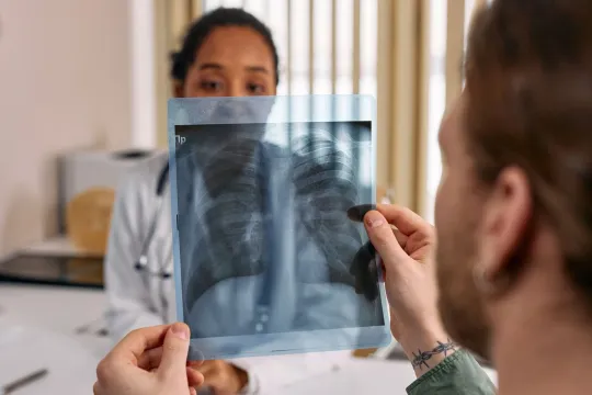
A chest X-ray is the most common imaging test, using a very small radiation dose to picture the heart and lungs. Learn why it is ordered, how it works, how to prepare, and how results are read.