Radiology: A Patient's Guide
The main imaging types, why scans are ordered, radiation safety, and what a radiologist's report does and does not tell you.

Radiology is the branch of medicine that uses imaging to see inside the body without surgery. If your doctor has ordered a scan and you want to understand what radiology actually is, why it is used, and what happens to the pictures afterward, this plain-language guide walks through the whole picture.
The short version
- Radiology covers X-ray, CT, MRI, ultrasound, and nuclear medicine: different tools for different questions.
- X-ray and CT use small amounts of ionizing radiation. Ultrasound and MRI do not use ionizing radiation at all.
- A radiologist, a doctor who specializes in imaging, reads your scan and writes a report for the doctor who ordered it.
- Images do not diagnose you by themselves. Your clinician combines them with your history, exam, and other tests.
What radiology means
Radiology is the medical specialty that creates and interprets pictures of the inside of the body to diagnose and sometimes treat disease. The images are produced by machines that pass energy through or around you, then reconstruct what that energy reveals about bone, soft tissue, fluid, and air. According to MedlinePlus, an X-ray, the oldest imaging tool, sends a small amount of radiation through your body to create pictures of bones and organs, with dense bone absorbing the most and appearing white while air-filled lungs appear black.
There are two broad branches worth knowing. Diagnostic radiology is about seeing and reporting: making the images and interpreting them. Interventional radiology uses the same imaging to guide minimally invasive procedures, such as placing a needle precisely or treating a blood vessel through a tiny incision, so that open surgery can sometimes be avoided. Most scans an average patient encounters fall under diagnostic radiology.
Modern radiology is much broader than a single X-ray film. It is an umbrella for several distinct technologies, and part of a good scan experience is having the right one ordered for your specific question. This article is the hub of a small cluster: for the deeper detail on any single modality, follow the internal links to guides such as the CT scan explainer and the MRI scan explainer.
The main types of imaging

Each technology answers a different kind of question. A referring doctor and radiologist choose based on the organ, the suspected problem, and safety.
- X-ray (radiography): fast, widely available, best for bones, the chest, and some abdominal problems. Uses a small dose of ionizing radiation.
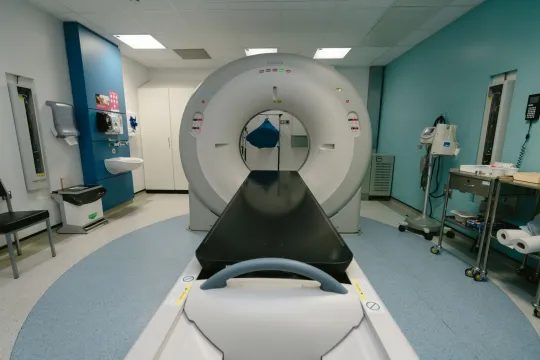
- CT (computed tomography): many X-ray images combined by computer into detailed cross-sections. Excellent for trauma, the chest, abdomen, and blood vessels. Uses more ionizing radiation than a plain X-ray.
- MRI (magnetic resonance imaging): uses a strong magnet and radio waves, no ionizing radiation. Superb for the brain, spine, joints, and soft tissue.
- Ultrasound (sonography): uses high-frequency sound waves, no ionizing radiation. Common for pregnancy, the abdomen, and blood vessels.
- Nuclear medicine and PET: a small amount of radioactive tracer highlights how organs or tissues are functioning, not just how they look.
Why a doctor orders imaging
Imaging is ordered to answer a specific clinical question, not to browse the body at random. Common reasons include finding the cause of pain, checking for a fracture, looking for signs of infection like pneumonia, screening for disease such as breast cancer with a mammogram, measuring bone strength with a DEXA scan, guiding a procedure, or monitoring a known condition over time. To keep imaging appropriate, professional bodies publish evidence-based guidance; the American College of Radiology, for example, maintains criteria that help clinicians pick the most suitable test for a given situation.
Appropriate use also protects you. Ordering the right test the first time can avoid a second scan, reduce unnecessary radiation, and get you an answer faster. If a test is repeated, it is often to compare against an earlier image and track change over time, which is why keeping copies of prior scans is genuinely useful.
How a radiology exam works

The exact steps depend on the modality, but the shape is similar. You check in, confirm your identity and the exam ordered, and remove metal or clothing over the area being imaged. A technologist positions you and operates the machine. For X-ray and CT you hold still, and sometimes hold your breath, while the images are captured. For MRI you lie inside the scanner while it makes loud knocking sounds. For ultrasound a technologist glides a probe over gel on your skin.
Some exams use a contrast agent, a substance given by mouth, injection, or other route, that makes certain tissues or vessels stand out. Tell the staff about allergies, kidney problems, pregnancy, or breastfeeding before any contrast is given.
Preparation tip
Ask the clinic when you book whether you need to fast, adjust medication, arrive early, or bring prior images. Wear simple clothing without metal, and bring a list of your current medications and any past scans of the same area so the radiologist can compare.
Is radiology safe? Radiation explained
Safety depends on the tool. Ultrasound and MRI do not use ionizing radiation. X-ray and CT do, but in carefully controlled amounts. RadiologyInfo, from the Radiological Society of North America and the ACR, notes that facilities follow the ALARA principle, meaning As Low As Reasonably Achievable, to keep doses down while still getting a diagnostic image. For context, a chest X-ray delivers roughly 0.1 millisieverts, comparable to about 10 days of the natural background radiation everyone receives, while a CT of the abdomen and pelvis is higher at around 7.7 millisieverts.
The guiding idea is that for an appropriate scan, the benefit of an accurate diagnosis outweighs the small radiation risk. If you are or might be pregnant, tell the team before any X-ray or CT, because doctors avoid unnecessary radiation to a fetus. MRI has its own safety rules centered on the strong magnet: metal implants and certain devices must be checked in advance.
What your results mean
After the exam, a radiologist reviews the images and writes a report describing the findings and an impression. That report goes to the doctor who ordered the test. Results may be ready the same day or take longer depending on the exam and whether prior images are compared. A finding on a scan is a piece of information, not a diagnosis on its own: the same appearance can have several causes, and only your clinician, who knows your symptoms and history, can interpret what it means for you.
Radiology reports use particular language that can look alarming out of context. Words like "unremarkable" are good news, meaning nothing abnormal was seen. An "incidental finding" is something spotted by chance that may be harmless and unrelated to why you were scanned. Terms such as "correlate clinically" are the radiologist's way of saying the images should be read together with your symptoms and other tests. If a phrase worries you, ask your doctor to explain it rather than searching it in isolation.
This is general information, not medical advice
Nothing here can tell you what your particular scan or symptom means. Always discuss your results, your risks, and your next steps with the doctor who ordered the imaging or with your radiologist. If you have an urgent or severe symptom, seek care right away rather than waiting for a scan.
Choosing where to get imaged
Where you go affects image quality and comfort. A practical checklist: is the facility accredited, does it offer the specific scan your doctor ordered, does it accept your insurance, and is a board-certified radiologist reading the images. Accreditation from a body like the American College of Radiology signals that equipment, staff, and quality controls have been independently reviewed. To compare options sensibly, see our honest guide on how to choose a medical imaging center, learn what ACR accreditation means, or start with a local search using our radiology near me guide.
Frequently asked questions
What is the difference between radiology and radiography?
Radiology is the whole medical specialty of imaging, including X-ray, CT, MRI, ultrasound, and nuclear medicine. Radiography specifically means producing images with X-rays, so it is one part of radiology. A radiologist is the doctor who interprets the images, while a radiographer or technologist operates the equipment.
Which radiology tests use radiation?
X-ray, CT, and nuclear medicine use ionizing radiation. Ultrasound and MRI do not use ionizing radiation at all. When radiation is used, facilities follow the ALARA principle to keep the dose as low as reasonably achievable while still getting a useful image.
Do I need a referral for a radiology scan?
In most systems a doctor must order the scan and provide the clinical reason, and many insurers require this before they will cover it. Some clinics offer certain self-pay scans without a referral, but the results still need a clinician to interpret them in the context of your health.
How long does it take to get results?
It varies. Some results are ready the same day, while others take several days, especially if the radiologist is comparing your scan with earlier images. Ask the clinic when and how you will get your report, and follow up with the doctor who ordered the test.
Can a radiology scan tell me my diagnosis?
Not by itself. A scan provides images and a radiologist's report, but the same finding can have different causes. Your diagnosis comes from a clinician who combines the imaging with your symptoms, history, exam, and any other tests. Always review results with your healthcare provider.
More guides on Ultrasound & Radiology (General)
Have a question or a topic to suggest?
We read every message. Tell us what you would like to see next.
You may also like

CT Scan: Uses, Procedure and Results
A CT scan, or CAT scan, uses x-rays and a computer to create detailed cross-sectional images of the body. Here is why doctors order one, how the procedure works, what contrast dye does, and how the radiation compares.

MRI Scan: Uses, Procedure & Results
A plain-language guide to MRI scans: what they show, why a doctor might order one, how to prepare, what happens during the scan, and how results are read.
Medical Imaging Centers Explained
What a center for medical imaging is, the services it offers, what happens at your visit, and how to choose an accredited, good-value one.