Pancreas Ultrasound
A pancreas ultrasound is part of a standard abdominal ultrasound that uses sound waves to picture the pancreas and the organs around it.

A pancreas ultrasound is part of a standard abdominal ultrasound that uses sound waves to picture the pancreas and the organs around it. It is safe, painless and uses no radiation, and doctors often order it as a first look when they are investigating upper abdominal pain, abnormal blood tests or a possible problem with the gland. This guide explains why the scan is ordered, how to prepare, what happens during it and how to make sense of the report.
The short version
- The pancreas is imaged as part of an abdominal ultrasound, using sound waves and no ionizing radiation.
- It is commonly ordered for upper abdominal pain, suspected gallstones or pancreatitis, and abnormal liver or pancreas blood tests.
- You are usually asked to fast for several hours so the view is not blocked by food or bowel gas; follow your clinic's instructions.
- The pancreas sits deep in the abdomen and can be hard to see, so a CT or MRI is often added. Only a radiologist and your doctor can interpret the images.
What a pancreas ultrasound is
The pancreas is a long, soft gland tucked behind the stomach that helps you digest food and control blood sugar. A pancreas ultrasound is not usually a stand alone test. Instead, the pancreas is examined as one organ within an abdominal ultrasound, which also views the liver, gallbladder, bile ducts, spleen, kidneys and the main abdominal blood vessel. As RadiologyInfo.org explains, an abdominal ultrasound evaluates the kidneys, liver, gallbladder, bile ducts, pancreas, spleen and abdominal aorta.
The scan works by sending high frequency sound waves from a small hand held probe, called a transducer, into the body. The probe records the echoes that bounce back from different tissues and a computer turns them into a live image on screen. Because it uses sound rather than radiation, ultrasound is safe, noninvasive and can be repeated as often as needed. For the technology in general, see our plain language ultrasound explainer.
What a pancreas ultrasound can show

Because the pancreas lies deep in the abdomen, ultrasound gives a useful but sometimes partial view. When the gland is seen clearly, the scan may help assess:
- Size and outline: whether the pancreas looks swollen, shrunken or normal.
- Signs of inflammation: changes that can accompany acute or long standing pancreatitis.
- Cysts and fluid collections: fluid filled areas in or near the gland.
- Ducts: whether the pancreatic or bile ducts look widened, which can point to a blockage.
- Nearby causes: gallstones in the gallbladder or bile ducts, which are a common trigger for pancreatitis.
Ultrasound is often the first step because it is quick and safe, but it is rarely the last word for the pancreas. When more detail is needed, your doctor may add a CT scan or an MRI, which see the whole gland more reliably. A pancreas check is usually part of a wider survey, so our guides to the liver ultrasound and the USG abdomen are worth reading alongside this one.
Why a doctor orders one
Clinicians reach for an abdominal ultrasound that includes the pancreas because it is safe, fast and gives helpful first line information. Common reasons include:
- Pain in the upper middle or upper right abdomen.
- Suspected gallstones or a suspected episode of pancreatitis.
- Abnormal blood tests, such as raised liver enzymes, amylase or lipase.
- Yellowing of the skin or eyes, which can signal a blocked bile duct.
- Following up a cyst or other finding seen on an earlier scan.
- Guiding a procedure such as a biopsy or drainage.
How to prepare

Preparation for a pancreas ultrasound is usually about clearing the view. Food in the stomach and gas in the bowel scatter sound waves and can hide the gland, so most clinics ask you to fast. RadiologyInfo.org notes that the exam requires little to no special preparation and that your doctor will tell you whether to refrain from eating or drinking beforehand. Typical steps include:
- Fast for about six to eight hours before the scan if your clinic asks you to, taking only small sips of water for essential medicines.
- Wear loose, comfortable clothing that lets you expose your abdomen easily.
- Bring your referral, a list of your medicines and any previous abdominal imaging.
- Leave jewelry at home and arrive in good time so you are not rushed.
Tip: If you have had abdominal imaging before, bring the earlier report or images. Comparing new pictures with older ones helps the radiologist judge whether a cyst or the size of the gland has changed over time.
What happens during the scan
An abdominal ultrasound that includes the pancreas usually takes about twenty to thirty minutes. You lie on a couch, most often on your back, and the sonographer spreads a warm water based gel over your upper abdomen so the sound waves pass in cleanly. They glide the probe across the skin, pressing gently and changing angle to reach behind the stomach, and may ask you to breathe in and hold your breath or to turn onto your side. These small movements shift the stomach and bowel out of the way and bring the pancreas into a better position.
The gel can feel cool at first and the probe may press over tender areas, but the scan is painless and involves no needles for a standard study. Afterward the gel is wiped off and, if you fasted, you can normally eat and return to your usual activities straight away.
Understanding your results
A radiologist reviews the recorded images and writes a report for the doctor who referred you. You usually will not get your results during the appointment, because the sonographer captures the pictures and the radiologist interprets them afterward, often within a few days.
Reports are descriptive. You might read that the pancreas appears normal in size and outline, that part of the gland could not be seen because of overlying bowel gas, that there is a cyst, or that the ducts look widened. These are observations, not a final diagnosis, and because the pancreas is a difficult organ to image, a normal or incomplete ultrasound does not always rule a problem in or out. That is why the same words can mean different things for different people, and why further tests are common.
Important: This article is general educational information, not medical advice, and it cannot tell you what your own pancreas scan means. Please discuss your report, symptoms and any next steps with your referring doctor or a radiologist.
Benefits, limits and safety
A pancreas ultrasound is safe, painless and uses no ionizing radiation, so it can be repeated freely and is suitable during pregnancy. It is widely available, comparatively inexpensive and gives real time images without needles or contrast for a standard study.
Its main limit is anatomy. The pancreas sits deep behind the stomach, and bowel gas or a larger body can block the sound waves so that part of the gland is not seen. Ultrasound is also operator dependent, meaning the skill of the sonographer matters. For these reasons a clear picture of the whole pancreas often needs a CT scan or an MRI, and a specialised test called endoscopic ultrasound can look at the gland from inside the digestive tract. These are decisions for your medical team based on your symptoms and earlier results.
How to find a good clinic
When you choose where to have your scan, put quality ahead of the lowest price. Confirm that the clinic is accredited, that scans are performed by qualified sonographers and reported by a radiologist, and that the report will reach your doctor promptly. Ask whether your images are stored so they can be compared with future scans. Our guide to how to find and compare ultrasound clinics walks through what to check before you book.
Prices vary by country, provider and whether you pay privately or use insurance, so confirm the fee, what it covers and your cover before booking. A scan bundled with a radiologist report generally offers better value than a cheaper scan with no interpretation.
Frequently asked questions
Is there a separate pancreas ultrasound, or is it part of an abdominal scan?
In most cases the pancreas is imaged as one organ within a standard abdominal ultrasound, which also views the liver, gallbladder, bile ducts, spleen, kidneys and aorta. There is not usually a separate pancreas only appointment.
Do I need to fast before a pancreas ultrasound?
Usually yes. Most clinics ask you to fast for several hours so food and bowel gas do not block the view of the gland. You can normally take small sips of water for essential medicines, but follow the exact instructions your clinic gives you.
Can an ultrasound see the whole pancreas?
Not always. The pancreas lies deep behind the stomach, so bowel gas or body size can hide part of it. When a full and detailed view is needed, doctors often add a CT scan or MRI, and sometimes an endoscopic ultrasound.
Is a pancreas ultrasound safe?
Yes. It uses sound waves rather than ionizing radiation, so it carries no known risk, is safe in pregnancy and can be repeated as needed. You may feel gentle pressure from the probe, but the scan is painless and needs no needles for a standard study.
When will I get my results?
Usually not on the day. A radiologist reviews the images and writes a report, so your written result often arrives a few days later through the doctor who referred you, who will explain what it means for you.
Sources and further reading:
- Abdominal Ultrasound, RadiologyInfo.org for what the scan images, including the pancreas, and why it uses no radiation.
- Ultrasound scan, NHS for how ultrasound works, its safety and preparation.
- Diagnostic Imaging, MedlinePlus (US National Library of Medicine) for how imaging tests fit together.
More guides on Ultrasound & Radiology (General)
Have a question or a topic to suggest?
We read every message. Tell us what you would like to see next.
You may also like
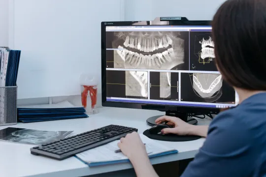
CT Imaging Near Me: Compare Clinics
Holding a CT referral and choosing between centers? Use a simple scorecard to compare accreditation, reporting, price, and turnaround instead of picking the first result.
DEXA Scan Near Me: Choose a Clinic
A DEXA scan is one test where the clinic you pick has a lasting effect, because results are compared over years. Learn how to choose a good bone density center.

Ultrasound Locations Near Me
Hospitals, imaging centers, and keepsake studios all advertise ultrasound. Learn to tell a proper diagnostic location from a novelty one before you book.