MRI: Uses, Procedure & Results
How a magnetic resonance image is made, what it shows, the metal safety rules, and what happens after your scan.

A magnetic resonance image, usually shortened to MRI, gives doctors an extraordinarily detailed view of soft tissue without using any radiation. This guide explains what an MRI is, why it is ordered, how the scan works, how to prepare, the safety rules around metal, and how your results are handled.
MRI can feel intimidating: the machine is large, the scan is noisy, and it takes longer than most other tests. Knowing what to expect removes most of that anxiety. Below we cover the whole process in plain language, along with the honest caveats about metal implants, claustrophobia, and what your report can and cannot tell you.
The short version
- MRI uses a strong magnetic field and radio waves, not ionizing radiation, to create detailed images.
- It excels at soft tissue: the brain, spinal cord, joints, muscles, and internal organs.
- Metal matters. Tell staff about any implants, devices, or metal fragments before your scan.
- A radiologist interprets the images and reports to your referring doctor, who explains what they mean for you.
What a magnetic resonance image is
MRI uses a powerful magnetic field, radiofrequency pulses, and a computer to produce detailed pictures of internal body structures. According to RadiologyInfo.org, the scanner uses a high-power magnet to temporarily change the position of hydrogen atoms that naturally exist within the body. As those atoms return to their normal state, they release energy that differs by tissue type, and a computer turns that signal into an image.
Because different soft tissues give off distinct signals, MRI can distinguish structures that look nearly identical on other scans. That soft-tissue contrast is its great strength. MRI is one of the major tools in radiology, sitting alongside X-ray, CT, and ultrasound, and each has situations where it is the best choice.
Why your doctor might order an MRI
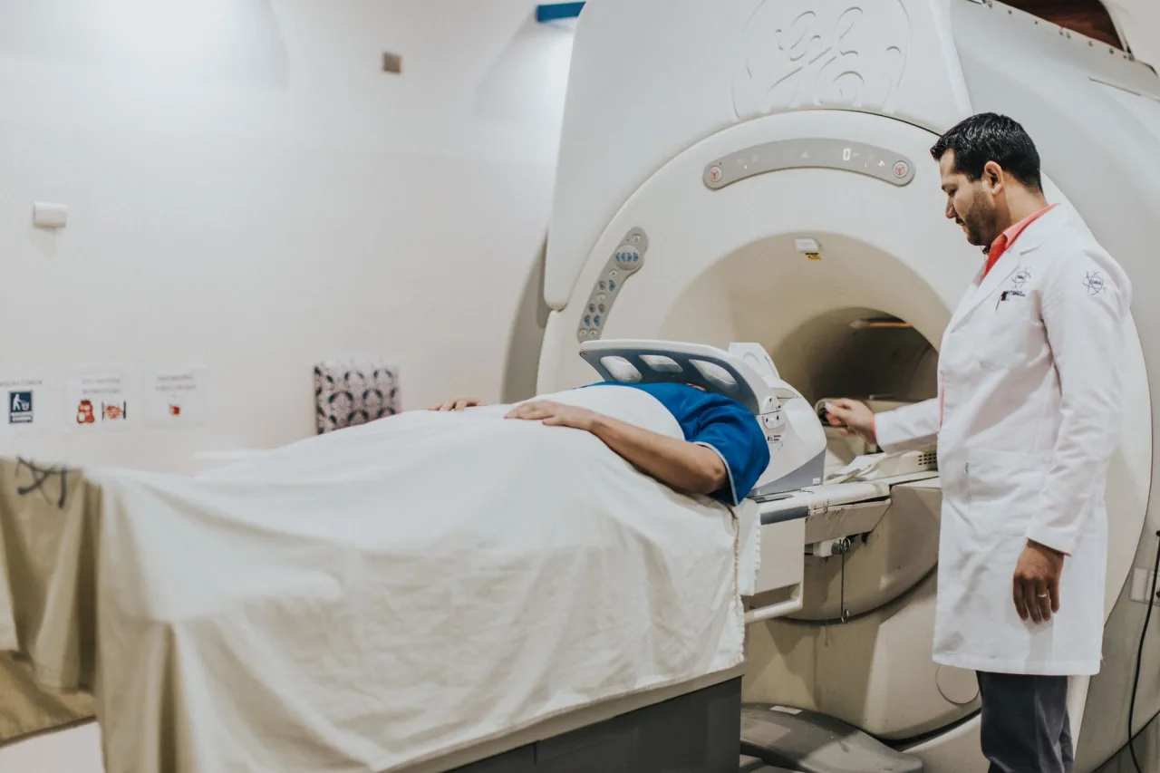
Doctors choose MRI when they need fine detail of soft tissue and want to avoid radiation. It is frequently used for the following.
- Brain and spine: stroke, tumours, multiple sclerosis, and disc or nerve problems.
- Joints and muscles: torn ligaments, cartilage damage, and sports injuries.
- Internal organs: the liver, kidneys, pancreas, and pelvic structures.
- Blood vessels: MR angiography can map vessels, sometimes without contrast.
- Cancer care: characterising a mass and planning treatment.
MRI and other scans are often complementary rather than interchangeable. Your doctor might start with an ultrasound or a CT scan and move to MRI for a clearer soft-tissue answer, or the other way around depending on the question.
How MRI compares with CT and X-ray
A common question is why one person gets an MRI and another gets a CT. The short answer is that they see different things. CT and X-ray use ionizing radiation and are fast and excellent for bone, bleeding, and lung detail. MRI uses no radiation and is superior for soft tissue such as the brain, spinal cord, and joints, but it is slower and more sensitive to movement.
- Radiation: MRI uses none, while CT and X-ray use ionizing radiation.
- Speed: CT takes minutes, MRI often takes 20 to 60 minutes.
- Best for: MRI for soft tissue, CT for bone, acute bleeding, and lung, ultrasound for real-time and pregnancy imaging.
How to prepare for an MRI
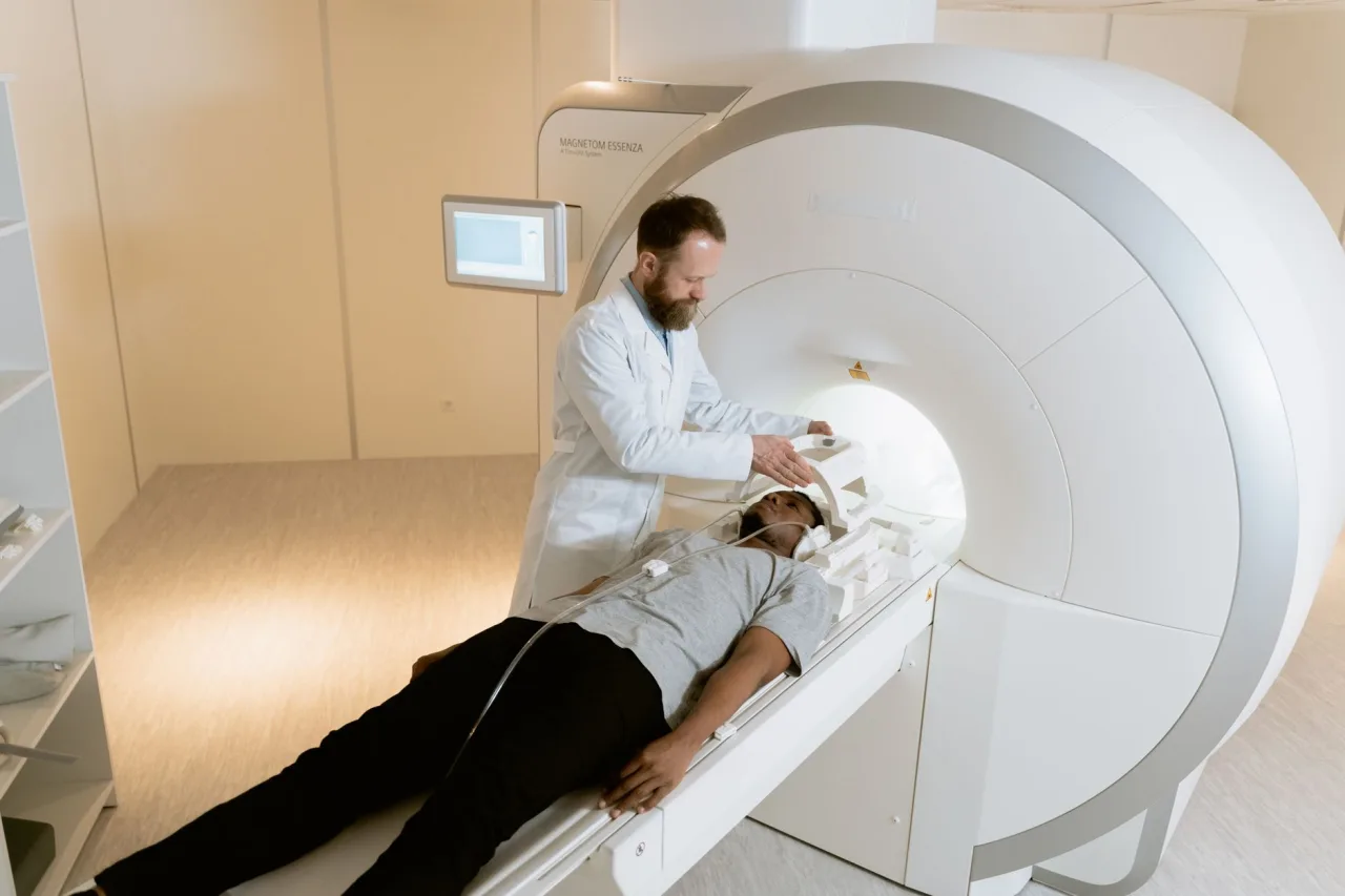
Preparation is mostly about safety and comfort. You will typically be asked to remove anything metallic, and to complete a detailed safety questionnaire. The NHS advises patients not to wear anything that has metal in it, such as jewellery, a bra with underwiring, hair clips, or hearing aids, and to tell staff about any metal parts inside the body such as surgical clips or a pacemaker.
- Metal and devices: declare implants, stents, clips, cochlear implants, pacemakers, and any history of metal fragments in the eyes.
- Clothing: you may change into a gown to avoid zips, hooks, and metallic threads.
- Eating: most MRIs need no fasting, but some abdominal scans do, so check your instructions.
- Contrast: if gadolinium contrast is planned, mention kidney problems, allergies, or pregnancy.
Important: The MRI magnet is always on, even between scans. Never take metal objects into the scan room, and always disclose implants and devices honestly. This is a genuine safety issue, not a formality.
What happens during the scan
You lie on a padded table that slides into a tube-shaped scanner. The machine makes loud knocking and buzzing sounds while it works, so you are given earplugs or headphones. Staff watch and talk to you from the next room, and you hold a buzzer to call them at any time. The most important thing you can do is stay still, because motion blurs the images.
Some scans use a contrast agent called gadolinium, injected into a vein to make certain tissues and vessels stand out. RadiologyInfo notes that gadolinium can be used safely in many patients who are allergic to iodine-based contrast, which is one reason MRI is sometimes preferred. A typical scan lasts from 20 to 60 minutes depending on the body part and the number of sequences.
During those sequences you may notice the noise changing in pitch and rhythm as the machine switches between different imaging steps. This is completely normal and simply reflects the scanner working through the set of images your doctor requested. For some body parts, such as the abdomen, you may also be asked to hold your breath briefly at certain points to keep the pictures sharp. Staff will explain each step and let you know roughly how long each part will last, so you are never left wondering what is happening.
Claustrophobia and the open MRI option
Lying still inside a narrow tube is difficult for some people. If enclosed spaces make you anxious, say so before the appointment. The NHS suggests talking to a healthcare professional beforehand, as you may be offered medicine to help you relax. Other strategies include going in feet first where possible, keeping your eyes closed, and using the provided music.
Wider or open-sided scanners can also help, though image detail can differ from a standard high-field machine. If an open design would make the difference between completing the scan or not, it is worth asking about. Our guide to finding open MRI options explains what to look for and the trade-offs to weigh.
Understanding your MRI results
After the scan, a radiologist studies the images and prepares a report for the doctor who referred you. The NHS notes it usually takes between one and two weeks to get MRI results, though urgent scans are read faster. You will normally discuss the findings with your referring doctor rather than the radiographer who ran the scan.
MRI reports are detailed and often list incidental findings that are common and harmless. A word such as "lesion," "signal change," or "degenerative" does not automatically mean something serious, and its significance depends on your symptoms and history. That is why interpretation belongs to your clinician, who sees the whole picture. Resist the urge to self-diagnose from the report wording alone.
Note: This article is general information, not medical advice. It cannot tell you what your own scan shows. Please review your MRI report and any symptoms with your referring doctor or a qualified radiologist.
Safety, limits, and typical costs
MRI is very safe for people without metal contraindications, and it avoids radiation entirely. Its main limits are practical: it is slower and noisier than other scans, less suited to acutely unwell patients who cannot lie still, and unavailable to some people with certain implants. Costs vary widely by country, provider, body region, and whether contrast is used. In public health systems a medically necessary MRI may be free at the point of care, while private scans can range from several hundred to over a thousand in local currency. These are broad ranges only, so confirm the fee with the imaging centre and check coverage with your insurer. When comparing facilities, our directory on choosing an MRI centre covers accreditation and quality signals to look for.
Frequently asked questions
Does an MRI use radiation?
No. An MRI uses a strong magnetic field and radio waves to create images, not ionizing radiation. This is a key difference from X-ray and CT scans, and it is one reason MRI is often chosen for repeated soft-tissue imaging.
Why can't I wear metal into the scanner?
The MRI magnet is extremely strong and always on. Metal objects can be pulled toward the machine, heat up, or distort the images, and some implanted devices can malfunction. Removing metal and declaring any implants keeps you safe and keeps the pictures clear.
How long does an MRI take?
Most MRI scans take between 20 and 60 minutes, depending on the body part and how many image sequences are needed. Scans that use contrast or cover several areas take longer. Staff will give you an estimate for your specific appointment.
What if I am claustrophobic?
Tell the clinic before your appointment. You may be offered medicine to help you relax, and strategies such as closing your eyes, going in feet first, or using an open-sided scanner can help. Staff monitor you throughout and you hold a buzzer to call them at any time.
When will I get my results?
A radiologist interprets the images and writes a report for your referring doctor, so you usually hear results from that doctor. It commonly takes about one to two weeks, though urgent scans are read sooner.
More guides on Ultrasound & Radiology (General)
Have a question or a topic to suggest?
We read every message. Tell us what you would like to see next.
You may also like

Ultrasound: Uses, Procedure & Results
An ultrasound uses sound waves, not radiation, to image soft tissue in real time. This guide covers why scans are ordered, how they work, how to prepare, and how results are interpreted, with a reminder to discuss findings with your doctor.

CT Scan: Uses, Procedure and Results
A CT scan, or CAT scan, uses x-rays and a computer to create detailed cross-sectional images of the body. Here is why doctors order one, how the procedure works, what contrast dye does, and how the radiation compares.
Chest X-Ray: Uses, Procedure & Results
A chest X-ray is the most common imaging test, using a very small radiation dose to picture the heart and lungs. Learn why it is ordered, how it works, how to prepare, and how results are read.