Abdominal Ultrasound: Uses and Results
Which organs it checks, why it is ordered, how to prepare, and how to read common findings.
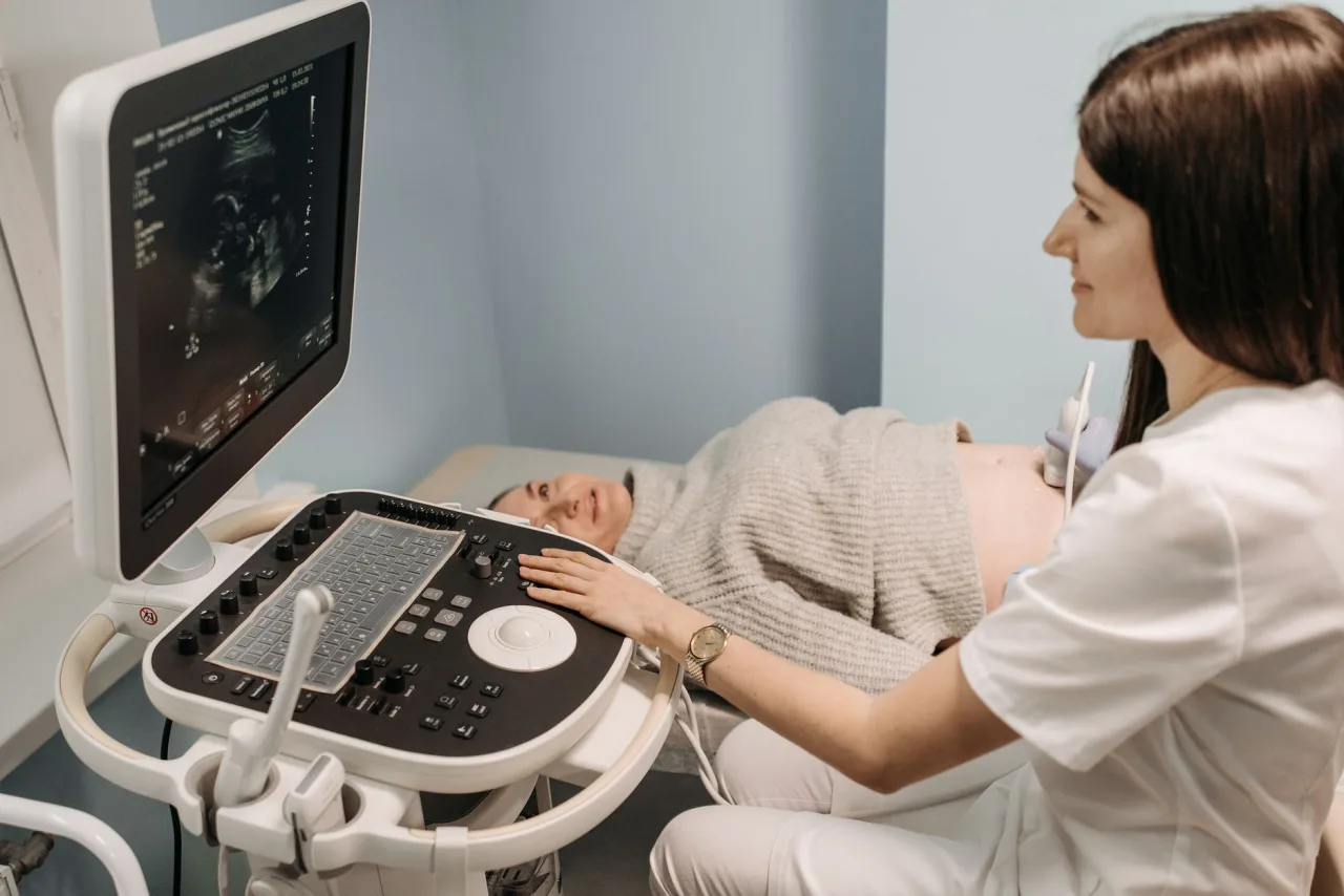
An abdominal ultrasound is a safe, radiation free scan that lets a doctor look at the organs inside your belly, including the liver, gallbladder, kidneys, pancreas and spleen. It is one of the most widely used imaging tests because it is quick, painless and gives a live view of how organs look and how blood flows. This guide explains why it is ordered, how to prepare, what happens during the scan and how to make sense of the report your doctor receives.
The short version
- An abdominal ultrasound pictures organs such as the liver, gallbladder, kidneys, pancreas, spleen and major blood vessels using sound waves.
- It is commonly ordered for belly pain, abnormal blood tests, suspected gallstones or kidney stones, and to check organ size.
- You are often asked to fast for several hours so the gallbladder is full and bowel gas is reduced for a clearer view.
- Only a radiologist and your doctor can interpret the images for you. A descriptive finding is not the same as a diagnosis.
What an abdominal ultrasound is
An abdominal ultrasound, also called an abdominal sonogram, uses high frequency sound waves to build a live moving image of the organs and blood vessels in your abdomen. A sonographer glides a small probe, the transducer, over the skin. The probe emits sound waves and records the echoes that return from tissue boundaries, and a computer turns those echoes into pictures on a screen in real time.
Unlike an X-ray or a CT scan, ultrasound uses no ionising radiation, which is a large part of why it is often the first imaging test for the abdomen. It shows soft tissue detail well, can watch organs move with breathing, and can measure blood flow when Doppler is added. For the broader picture of the technology, see our explainer on what an ultrasound is and how it is used.
What it can look at
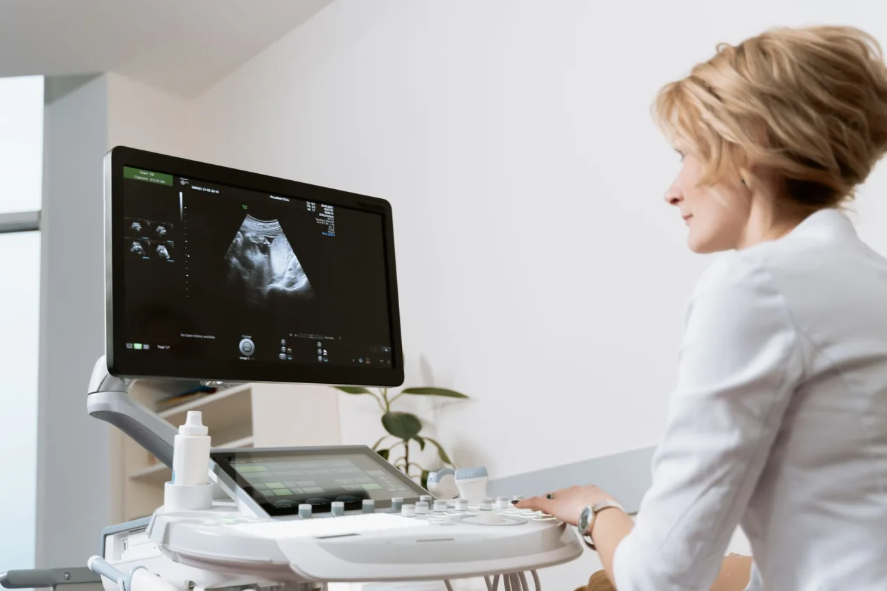
A full abdominal ultrasound typically surveys several organs in one sitting. Depending on the reason for your scan, the sonographer may assess:
- Liver: size, texture and signs of fatty change or lesions. A focused version of this is the liver ultrasound.
- Gallbladder and bile ducts: gallstones, wall thickening and blockage.
- Kidneys: size, stones, cysts, swelling and signs of obstruction.
- Pancreas and spleen: size and obvious abnormalities, though these can be partly hidden by bowel gas.
- Major blood vessels: the aorta and other vessels, for example to screen for an aneurysm.
When the scan is extended to the lower abdomen and pelvis it is often described separately. Our guide to the USG abdomen and pelvis covers that combined study.
Why a doctor orders one
Clinicians reach for abdominal ultrasound often because it is safe, fast and informative. Typical reasons include:
- Investigating the source of abdominal pain.
- Looking for gallstones or kidney stones.
- Following up abnormal blood tests, such as raised liver enzymes or kidney markers.
- Checking for organ enlargement, cysts, tumours or fluid in the abdomen.
- Screening the aorta for an aneurysm in people at risk.
- Guiding procedures such as draining fluid or taking a biopsy.
Ultrasound and cross sectional imaging often work together. If a finding needs more detail, your doctor may follow up with a CT scan. Each test has strengths, and the choice depends on what needs to be answered.
How to prepare
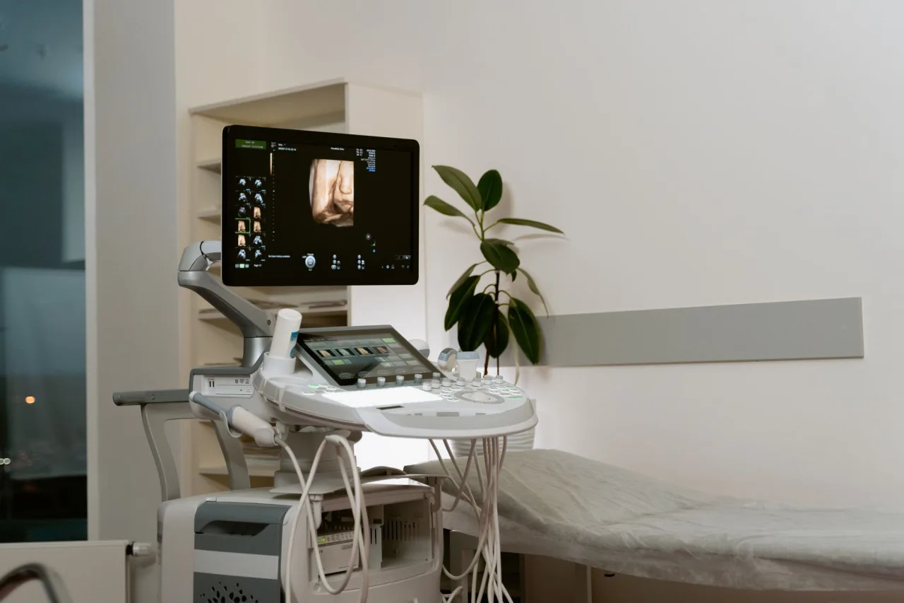
Preparation depends on the reason for the scan, so the single most important step is to follow the instructions your clinic gives you. For many abdominal ultrasounds, particularly when the gallbladder is a focus, you will be asked not to eat for several hours beforehand. Fasting keeps the gallbladder full and reduces bowel gas, both of which improve the view. For some kidney or pelvic questions you may instead be asked to drink water and arrive with a full bladder.
- Wear loose clothing and expect to change into a gown and uncover your abdomen.
- Bring your referral, previous imaging and a list of medicines.
- Ask in advance whether to fast, for how long, and whether to take your usual medication with water.
- Allow extra time for check in so you are relaxed for the scan.
Tip: If you have had a previous abdominal scan, bring the report or images. Comparing new pictures with older ones helps the radiologist judge whether anything has genuinely changed.
What happens during the scan
An abdominal ultrasound usually takes about twenty to thirty minutes. You lie on your back on a couch, and the sonographer applies a warm water based gel to your abdomen so the sound waves pass in cleanly. They move the probe across the skin, pressing gently and changing angle to view each organ, and may ask you to breathe in and hold your breath so organs drop into a better position.
The gel can feel cool at first and the probe may press firmly over tender areas, but the test is not painful and involves no needles for a standard scan. When it is done the gel is wiped off and you can normally return to eating and your usual activities immediately, unless you were told otherwise.
Understanding your results
A radiologist reviews the captured images and writes a report for the doctor who referred you. You often will not receive results during the appointment, because the sonographer records the pictures and the radiologist interprets them afterwards, usually within a few days.
Reports are descriptive. You might read that organs are normal in size and appearance, that there are gallstones, a simple kidney cyst, increased liver echogenicity suggesting fat, or a lesion that needs further assessment. These descriptions are observations, not a final verdict. Many findings, such as small simple cysts, are common and harmless, while others need context from your history and blood tests. That is why the same words can carry very different weight for two different people.
Important: This article gives general educational information and is not medical advice. It cannot tell you what your own results mean. Please discuss your report, symptoms and any next steps with your referring doctor or a radiologist.
Benefits, limits and safety
Abdominal ultrasound is safe, painless and uses no ionising radiation, so it can be repeated freely and is suitable during pregnancy. It is comparatively inexpensive, widely available and gives real time images with good soft tissue detail.
Its main limits come from physics. Sound waves are blocked by gas and bone and are weakened as they pass through more tissue, so bowel gas and a larger body habitus can reduce image quality, and deep structures such as the pancreas are not always fully seen. Ultrasound is operator dependent, meaning results rely on the skill of the sonographer, which is a good reason to choose an accredited clinic. When ultrasound cannot answer the question, a CT or MRI may be added.
How to find a good clinic
To choose where to have your scan, prioritise quality over the lowest price. Confirm that the clinic is accredited, that scans are performed by qualified sonographers and reported by a radiologist, and that the report will reach your doctor quickly. Ask whether your images are stored for future comparison. Our guide to how to find and compare ultrasound clinics walks through what to check before booking.
Prices vary by country, provider and whether you pay privately or use insurance, so confirm the fee, what it includes and your coverage before booking. A scan bundled with a radiologist report generally offers better value than a cheaper scan with no interpretation.
Frequently asked questions
Do I have to fast before an abdominal ultrasound?
Often yes, especially when the gallbladder is being examined. Many clinics ask you to avoid food for several hours so the gallbladder is full and bowel gas is reduced. Some kidney or pelvic scans instead ask for a full bladder. Always follow the specific instructions your clinic provides.
What organs does an abdominal ultrasound show?
It commonly examines the liver, gallbladder and bile ducts, kidneys, pancreas, spleen and major blood vessels such as the aorta. The exact organs assessed depend on the reason your doctor ordered the scan.
Is an abdominal ultrasound painful or risky?
No. The scan is painless and uses sound waves rather than ionising radiation, so it carries no known risk and can be repeated as needed. You may feel gentle pressure from the probe, especially over a tender area.
How long does the scan take and when will I get results?
The scan itself usually takes about twenty to thirty minutes. A radiologist then reviews the images and writes a report, so your written result often arrives a few days later through your referring doctor.
Can an abdominal ultrasound miss something?
It can. Bowel gas, body size and the depth of an organ can limit the view, and some findings are subtle. If a question remains, your doctor may add a CT or MRI. Discuss any concerns and your results with your clinician.
Sources and further reading:
- Abdominal Ultrasound, RadiologyInfo.org for how the scan works, common uses and safety.
- Abdominal Ultrasound, MedlinePlus (US National Library of Medicine) for preparation and the organs examined.
- Ultrasound scan, NHS for ultrasound types and why it uses no radiation.
More guides on Ultrasound Types & Procedures
Have a question or a topic to suggest?
We read every message. Tell us what you would like to see next.
You may also like
Liver Ultrasound: Uses and Results
A plain guide to the liver ultrasound: why doctors order it, how to prepare, what happens during the scan and how to understand the report your clinician receives.

USG Abdomen & Pelvis: Uses & Results
USG abdomen and pelvis is a common, radiation-free scan. Learn why it is ordered, how it works, how to prepare, and what the report language generally means.
Ultrasound: Uses, Procedure & Results
An ultrasound uses sound waves, not radiation, to image soft tissue in real time. This guide covers why scans are ordered, how they work, how to prepare, and how results are interpreted, with a reminder to discuss findings with your doctor.