Chest X-Ray: A Patient's Guide
What a chest X-ray shows, how it is performed, how safe it is, and why a normal result does not always rule everything out.

Chest radiology means imaging of the chest, and the chest X-ray is its most common exam. If your doctor has ordered one for a cough, chest pain, or shortness of breath, this guide explains what a chest X-ray shows, how the scan is done, whether it is safe, and how to make sense of the report.
The short version
- A chest X-ray is the most commonly performed diagnostic X-ray exam, imaging the heart, lungs, airways, and chest bones.
- It uses a small dose of ionizing radiation, about 0.1 millisieverts, roughly 10 days of natural background radiation.
- The exam is quick, painless, and usually needs no special preparation, but tell staff if you might be pregnant.
- A chest X-ray cannot rule out every chest problem; some conditions need CT or other tests to detect.
What chest radiology covers
Chest radiology is the imaging of the structures inside the chest, or thorax: the lungs, heart, airways, major blood vessels, and the bones of the chest wall and spine. The workhorse of chest radiology is the chest X-ray, also called chest radiography, which RadiologyInfo describes as the most commonly performed diagnostic X-ray examination. When a plain X-ray is not enough, chest radiology also includes CT of the chest and other studies. This guide focuses on the chest X-ray, the test most people will encounter first, and sits alongside our closely related chest radiography guide.
Why a chest X-ray is ordered
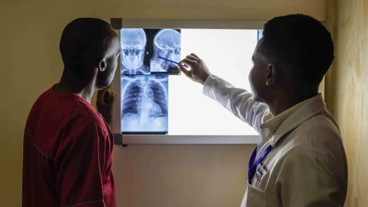
Doctors use chest X-rays to investigate symptoms and to monitor known conditions. According to RadiologyInfo, physicians order them to evaluate symptoms such as breathing difficulty, a persistent cough, chest pain or injury, and fever. A chest X-ray can help diagnose or monitor conditions including:
- Pneumonia and other lung infections
- Heart failure and an enlarged heart
- Emphysema and other chronic lung disease
- Lung cancer or other masses
- Fluid or air collecting around the lungs
It is also used to check the position of devices such as pacemakers or tubes, and to see how a known condition is responding to treatment. For the bigger picture of how this fits with other scans, see our radiology overview.
How the procedure works
A chest X-ray is straightforward. A technologist typically captures two views, one from the back and one from the side, while you stand against an imaging plate. RadiologyInfo notes the whole process takes about 15 minutes. You will be asked to remain still and to hold your breath briefly, because breathing blurs the image. The exam is painless, though the equipment or the required position can feel slightly uncomfortable for a moment.
The two standard views have names you may see on your report. The PA view, meaning posterior-anterior, is taken with the X-ray beam passing from your back to the front while you stand facing the plate. The lateral view is taken from the side. Together they let the radiologist judge structures in three dimensions and locate anything unusual. If you cannot stand, for example in a hospital bed, a single portable AP view may be taken instead, which is why a radiologist sometimes notes that image quality was limited by the technique available.
The NHS adds that you should remove metal items over the chest, and that the X-ray itself takes only a few seconds of stillness while the machine is lined up to the area being imaged.
Preparation tip
No special preparation is usually needed for a chest X-ray. Wear a top without metal buttons, zips, or jewelry, and be ready to change into a gown. Tell the staff before the scan if you are or might be pregnant so they can weigh alternatives.
Is a chest X-ray safe? Radiation in context
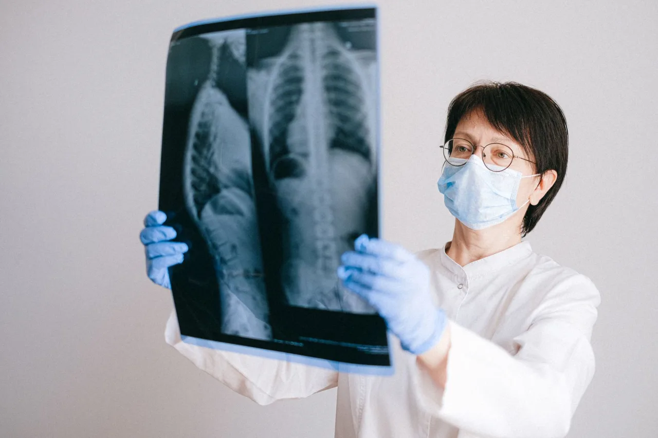
A chest X-ray uses a small amount of ionizing radiation. RadiologyInfo puts a chest X-ray at roughly 0.1 millisieverts, comparable to about 10 days of the natural background radiation everyone receives from the environment. MedlinePlus makes the same comparison, describing the exposure as about the same as the natural background over roughly 10 days. Facilities follow the ALARA principle, As Low As Reasonably Achievable, to keep the dose as low as possible while still producing a diagnostic image.
The NHS notes that X-rays are usually very safe and serious complications are rare, while acknowledging a very small risk from radiation with repeated exposure, which doctors weigh against the benefit of an accurate diagnosis. If you are pregnant, tell your care team before the scan, because doctors avoid unnecessary radiation to a fetus.
What the results mean
A radiologist reviews your chest X-ray and writes a report describing the findings and an impression, then sends it to the doctor who ordered the test. Results may be available the same day or within a few days. It is important to understand a key limitation: RadiologyInfo cautions that a chest X-ray cannot necessarily rule out all problems in the chest, because some conditions, such as a pulmonary embolism or a small cancer, may not show up on a conventional chest X-ray. That is why a normal chest X-ray does not always mean nothing is wrong, and why your doctor may order further tests such as a CT scan.
You may also come across common report phrases. A note that the lungs are "clear" and the heart size is "normal" is reassuring. A mention of an "opacity", "infiltrate", or "consolidation" points to an area that is denser than expected, which can reflect infection, fluid, or other causes, and needs a clinician to interpret. A "nodule" is a small spot that may be followed up to make sure it is stable. None of these words is a diagnosis by itself, and their meaning depends entirely on your symptoms and history, so let your doctor explain rather than drawing conclusions from the wording alone.
This is general information, not medical advice
This guide explains chest X-rays in general terms. It cannot tell you what your specific images or symptoms mean. Always review your results and next steps with the doctor who ordered the scan or with your radiologist. If you have severe chest pain, sudden breathlessness, or another emergency symptom, seek urgent care immediately rather than waiting for a scan.
Chest X-ray versus chest CT
A chest X-ray is fast, low-dose, and widely available, which makes it a sensible first look. A chest CT combines many X-ray images into detailed cross-sections and can reveal small nodules, blood clots, and subtle changes that a plain X-ray misses, at the cost of a higher radiation dose. Your doctor chooses based on the question being asked. Neither is better in the abstract; each is the right tool for different situations. A common pattern is a chest X-ray first, followed by a chest CT only if the X-ray is inconclusive or a more detailed look is needed. This staged approach keeps radiation as low as reasonably achievable while still getting the answer.
Where to get a chest X-ray
Chest X-rays are available at hospitals, urgent care, and outpatient imaging centers. When choosing, look for accreditation, confirm the facility performs the exam your doctor ordered, check insurance or self-pay cost, and make sure a board-certified radiologist reads the images. Our guide on how to choose a medical imaging center and our X-ray near me guide can help you compare options honestly.
Chest X-rays are among the most widely available imaging exams, so you usually have several options. If your doctor is monitoring a condition over time, it helps to return to the same facility, or at least to bring your prior images, so the radiologist can compare like with like and spot subtle change. If you are paying yourself, a chest X-ray is typically one of the more affordable studies, though prices still vary by country, region, and provider, so confirm the cost with the clinic and coverage with your insurer beforehand.
Frequently asked questions
How long does a chest X-ray take?
The whole process typically takes about 15 minutes, and the actual image capture takes only a few seconds of standing still and holding your breath. Usually two views are taken, one from the back and one from the side.
Is the radiation from a chest X-ray dangerous?
A chest X-ray uses a very small dose, about 0.1 millisieverts, comparable to roughly 10 days of natural background radiation. It is generally considered very safe, and facilities keep the dose as low as reasonably achievable. Tell your care team if you are or might be pregnant.
Do I need to prepare for a chest X-ray?
No special preparation is usually required. You will remove metal objects and jewelry over the chest and may change into a gown. Let staff know beforehand if you could be pregnant so they can consider alternatives.
Can a chest X-ray miss something?
Yes. A chest X-ray cannot rule out all chest problems, and some conditions such as a pulmonary embolism or a small cancer may not appear on it. A normal result does not always mean nothing is wrong, so your doctor may order further tests like a CT scan.
Who interprets my chest X-ray?
A radiologist, a doctor who specializes in imaging, reviews the images and writes a report for the doctor who ordered the exam. That referring doctor then discusses the findings with you in the context of your symptoms and history.
More guides on Ultrasound & Radiology (General)
Have a question or a topic to suggest?
We read every message. Tell us what you would like to see next.
You may also like

Radiology: A Patient's Guide
What radiology is in plain language: the main imaging types, why doctors order scans, how they work, radiation safety, and how to read your results.
Chest X-Ray: Uses, Procedure & Results
A chest X-ray is the most common imaging test, using a very small radiation dose to picture the heart and lungs. Learn why it is ordered, how it works, how to prepare, and how results are read.

CT Scan: Uses, Procedure and Results
A CT scan, or CAT scan, uses x-rays and a computer to create detailed cross-sectional images of the body. Here is why doctors order one, how the procedure works, what contrast dye does, and how the radiation compares.