Ultrasound: Uses, Procedure & Results
A plain-language guide to how ultrasound works, what it can see, how to prepare, and why your doctor, not the sonographer, explains your results.
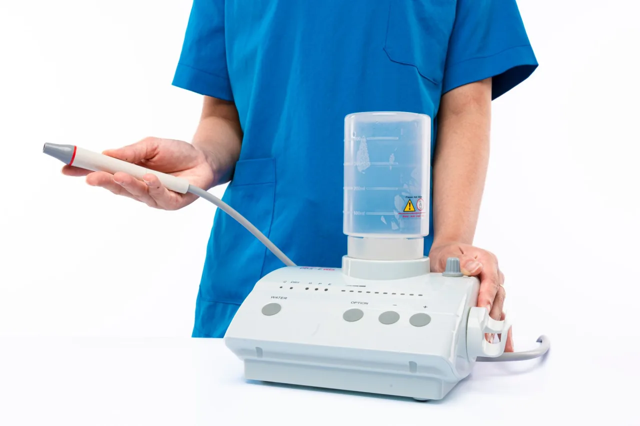
An ultrasound is one of the most common imaging tests in medicine, and for good reason: it is quick, painless, and uses no radiation at all. This guide explains what an ultrasound is, why doctors order one, how the scan actually works, how to prepare, and what happens after the images are taken.
If your clinician has referred you for an ultrasound, you probably have questions. What will it feel like? Do you need to fast or drink water first? What can the scan see, and what does it mean when the sonographer goes quiet and keeps measuring? This article walks through the whole journey in plain language, so you arrive informed and leave your appointment able to ask better questions.
The short version
- Ultrasound uses high-frequency sound waves, not radiation, so it is considered very safe and is the go-to scan in pregnancy.
- It is used to look at soft tissues and organs such as the liver, kidneys, heart, blood vessels, and a developing baby.
- Preparation depends on the exam: some need a full bladder, some need fasting, and many need none at all.
- The sonographer captures images, but a radiologist or your referring doctor interprets them and gives you the results.
What an ultrasound actually is
Ultrasound imaging uses sound waves to produce pictures of the inside of the body. A small handheld device called a transducer sends pulses of high-frequency sound into your tissues and then listens for the echoes that bounce back. A computer converts those echoes into a moving image on a screen in real time. Because it captures motion, ultrasound is especially good at showing things that move, such as blood flowing through a vessel or a heart valve opening and closing.
The single most important safety point is that ultrasound does not use ionizing radiation. According to RadiologyInfo.org, ultrasound is safe, noninvasive, and does not use radiation, and standard diagnostic ultrasound has no known harmful effects on humans. That is why it is the first-choice imaging test during pregnancy and for many everyday abdominal and pelvic questions. Ultrasound is one branch of the wider field of radiology, which also includes X-ray, CT, and MRI.
Why your doctor might order one

Doctors reach for ultrasound whenever they want a live look at soft tissue without exposing you to radiation. The NHS notes that ultrasound scans do not use any radiation, unlike X-rays or CT scans, which makes them a low-risk first step for a wide range of questions.
- Investigating symptoms: pain, swelling, or signs of infection in the abdomen or pelvis.
- Checking organs: the liver, gallbladder, kidneys, pancreas, spleen, bladder, thyroid, ovaries, and uterus.
- Assessing the heart and vessels: an echocardiogram looks at heart structure, and Doppler ultrasound measures blood flow and can find clots or narrowing.
- Pregnancy care: confirming a pregnancy, dating it, and monitoring the baby's growth and wellbeing.
- Guiding procedures: steering a needle accurately during a biopsy or fluid drainage.
Different body areas often have their own focused scan. Common examples include an abdominal ultrasound for the digestive organs and a transvaginal ultrasound for a closer look at the female pelvic organs.
The main types of ultrasound
Not every ultrasound is done the same way. The approach depends on how close the probe needs to be to the target tissue.
- External: the transducer is moved across the skin over the area of interest, after a layer of gel is applied. This is the familiar pregnancy and abdominal scan.
- Internal: a slim probe is gently inserted into a body cavity, such as the vagina or rectum, to sit nearer the organs being examined.
- Endoscopic: a tiny ultrasound probe is mounted on a flexible endoscope and passed into the digestive tract to image from the inside. This specialised technique is explained further in our endoscopic ultrasonography guide.
- Doppler and 3D or 4D: Doppler adds colour to show blood flow, while 3D and 4D reconstruct volume images, often used in obstetrics.
How to prepare for your scan

Preparation is exam-specific, and following the instructions your clinic gives you matters more than any general rule. That said, a few patterns are common.
- Full bladder: for many lower abdominal and early pregnancy scans, you may be asked to drink water beforehand and not empty your bladder, because a full bladder pushes the bowel aside and creates a clearer window.
- Fasting: for gallbladder and upper abdominal scans, you may be asked not to eat for several hours, so the gallbladder stays full and easy to see.
- No preparation: many scans, such as those of the thyroid, muscles, or blood vessels, need nothing special.
Wear loose, comfortable clothing and leave valuables at home, since you may be asked to remove jewellery from the area being scanned. If you are unsure whether to eat, drink, or take your usual medications, call the clinic and ask before the day.
Tip: Always confirm your preparation instructions when you book. A scan done with an empty bladder when a full one was needed sometimes has to be repeated, which costs you another trip.
What happens during the appointment
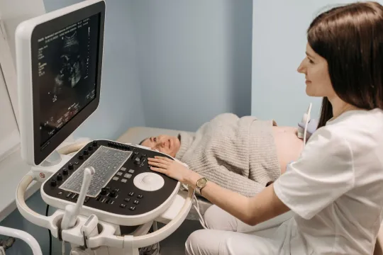
A typical external ultrasound is straightforward and comfortable. You lie on a couch, and the sonographer applies a warm water-based gel to your skin. The gel removes air between the probe and your body, because sound waves do not travel well through air. The sonographer then moves the transducer over the area, pressing gently and angling it to capture different views. You will often see a live black-and-white image on the monitor.
The scan is usually painless, though a probe pressed over a tender area can feel uncomfortable. Most appointments last between 15 and 45 minutes depending on how much needs to be examined. It is normal for the sonographer to pause, take measurements, and save still images without talking through every one. They are gathering the pictures a radiologist will later interpret, and they are often not permitted to give you a diagnosis on the spot.
Understanding your results
Here is the honest truth about results: the image itself is only half the story, and interpreting it is a skilled medical judgment. After your scan, a radiologist reviews the images and writes a report, which goes back to the doctor who referred you. Depending on the clinic and the reason for the scan, you might hear back the same day or wait a week or two.
Terms in a report such as "cyst," "shadowing," "echogenic," or "incidental finding" can sound alarming out of context, but many findings are benign or need only routine follow-up. What a result means for you depends on your symptoms, history, and other tests, which is exactly why interpretation belongs with your clinician rather than an internet search. If a finding needs a closer look, your doctor may order a follow-up scan or a different test such as an MRI.
Important: This article is general information, not medical advice, and it cannot tell you what your own results mean. Always discuss your report and any symptoms with your referring doctor or a qualified radiologist.
How safe is ultrasound, really?
Ultrasound has an excellent safety record, and its lack of ionizing radiation is a genuine advantage over X-ray and CT. There are no known harmful effects from standard diagnostic ultrasound at the levels used in clinics. The main practical limitation is not safety but physics: sound waves cannot pass well through bone or air, so ultrasound is less useful for imaging the lungs, the adult brain, or gas-filled bowel. For those areas, doctors turn to other scans.
One responsible caution: keepsake or "boutique" pregnancy scans done purely for souvenir images, without a medical reason, are discouraged by regulators because there is no clinical benefit to justify even a very low theoretical exposure. When ultrasound is ordered by your clinician for a medical reason, the benefit clearly outweighs any concern. If you are choosing where to have a medically indicated scan, our directory of how to find and compare ultrasound clinics walks through what to look for.
What an ultrasound typically costs
Cost varies widely by country, provider, body part, and whether you have insurance or a public health service. In many public systems a medically necessary ultrasound is free at the point of care. Where you pay privately, a straightforward scan often falls somewhere in the low hundreds in local currency, while specialised or contrast-enhanced studies cost more. These are broad ranges only. Prices and coverage differ enormously between providers and insurers, so confirm the exact fee with the clinic and check what your insurer covers before you book.
Frequently asked questions
Does an ultrasound hurt?
No, an ultrasound is generally painless. You feel the smooth probe and cool or warm gel moving across your skin, and at most mild pressure. If the probe passes over a tender or inflamed area it can feel briefly uncomfortable, but the scan itself does not cause pain.
Is ultrasound safe during pregnancy?
Yes. Ultrasound uses sound waves rather than radiation, which is why it is the standard imaging test in pregnancy and has no known harmful effects at diagnostic levels. Non-medical keepsake scans are discouraged, but scans ordered by your clinician for a medical reason are considered safe.
Do I need to fast or drink water before my scan?
It depends on the exam. Upper abdominal and gallbladder scans often require fasting, while lower abdominal and early pregnancy scans often require a full bladder. Many other scans need no preparation at all. Always follow the specific instructions your clinic gives you when you book.
When will I get my results?
A radiologist interprets the images and sends a report to your referring doctor, so you usually receive results from that doctor rather than the sonographer. Depending on the clinic and urgency, this can take from the same day to a week or two.
Why can't the sonographer tell me what they see?
Sonographers capture the images, but a radiologist or your doctor is responsible for interpreting them and giving a diagnosis. In many settings sonographers are not permitted to discuss findings, which is a normal part of the process and not a sign that something is wrong.
More guides on Ultrasound & Radiology (General)
Have a question or a topic to suggest?
We read every message. Tell us what you would like to see next.
You may also like

MRI: Uses, Procedure & Results
MRI uses a strong magnetic field and radio waves, not radiation, to produce detailed soft-tissue images. Learn why MRIs are ordered, how the scan works, the metal safety rules, and how a radiologist interprets your results.
Abdominal Ultrasound: Uses and Results
A clear guide to the abdominal ultrasound: which organs it checks, why doctors order it, how to prepare and how to understand the report your clinician receives.

Sonography: 2026 Career Guide
Diagnostic medical sonographers capture the ultrasound images doctors rely on. This 2026 guide covers the education and certification paths, realistic salary ranges, job outlook, and how to decide if the career fits you.