Imaging
When a doctor says you need "imaging," they mean one of a handful of tests that take pictures of the inside of your body: an X-ray, ultrasound, CT
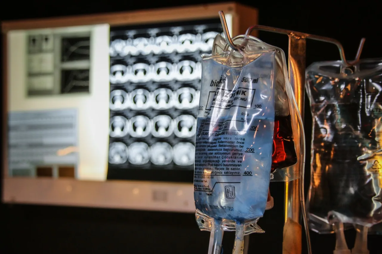
When a doctor says you need "imaging," they mean one of a handful of tests that take pictures of the inside of your body: an X-ray, ultrasound, CT, MRI, mammogram or nuclear scan. This hub explains why imaging gets ordered, what the whole journey looks like from referral to report, what is safe, and how to talk about your results with the right person.
The short version
- Imaging is a set of tools, not one test. Your doctor picks the one that best answers the specific question they have about your health.
- Ultrasound and MRI use no ionizing radiation. X-ray, CT and mammography use small, carefully controlled doses.
- Preparation depends entirely on the test: some need fasting or a full bladder, many need nothing. Follow the clinic's instructions exactly.
- A radiologist reads your images and writes a report for the doctor who ordered the scan. That doctor is the right person to explain what it means for you.
What "imaging" means and why a doctor orders it
Imaging, also called diagnostic imaging or radiology, is a group of techniques that let clinicians see structures and sometimes function inside the body without surgery. According to MedlinePlus, doctors use it to find health problems early, learn what is causing pain or other symptoms, plan treatment, and monitor a condition or check how well treatment is working. In other words, imaging is rarely done for its own sake: it is ordered to answer a defined clinical question.
That is the single most useful idea to carry into any appointment. There is no generic "scan" that shows everything. Sound waves see soft tissue and fluid well but cannot travel through bone or air. X-rays show bone and dense tissue clearly but give less detail in soft organs. A magnet reveals the brain, spine and joints in exquisite detail but is sensitive to metal. Your doctor weighs the question against these strengths and blind spots, then chooses. For a side-by-side comparison of the tools, our overview of medical imaging goes deeper on each modality.
The main types of imaging at a glance
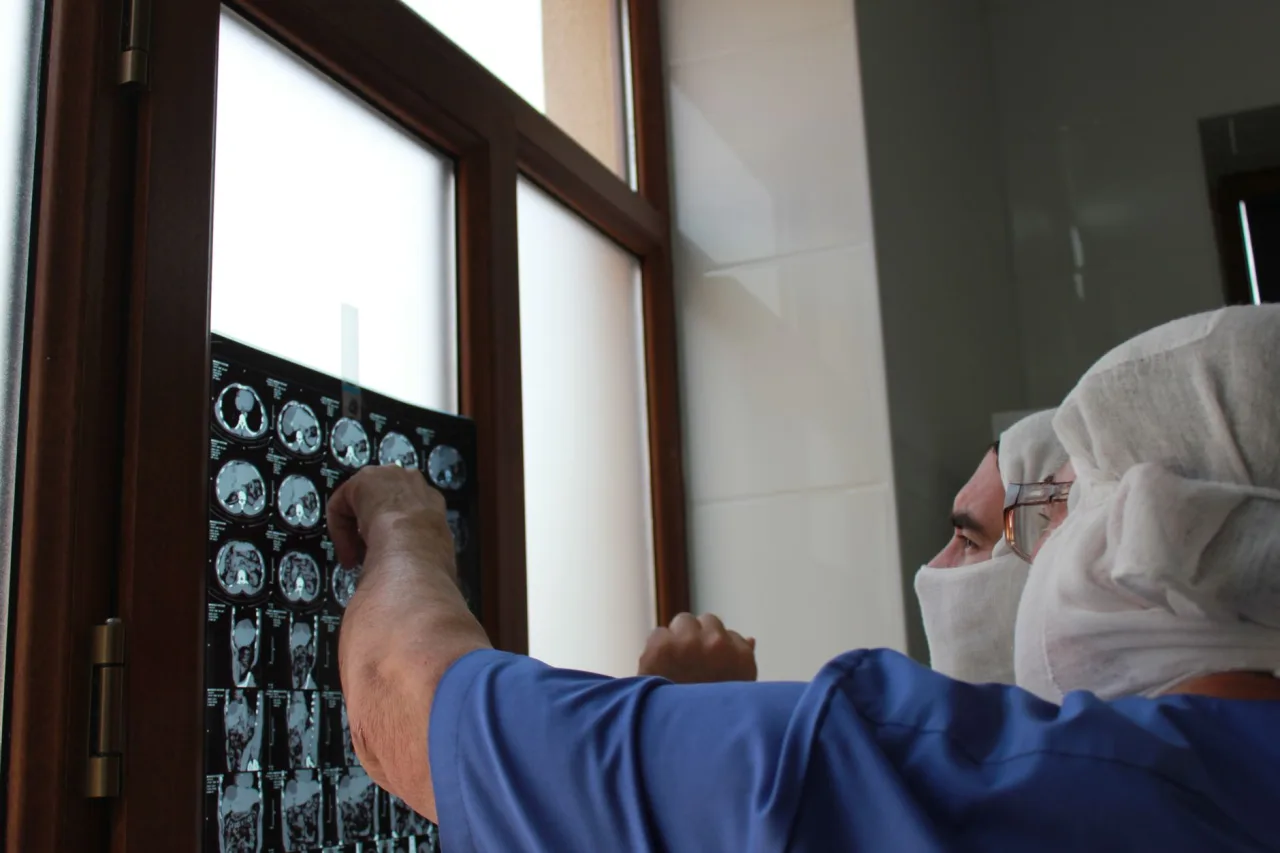
Here is how the common methods differ in everyday practice:
- X-ray: fast, inexpensive and widely available. Best for bones, the chest and dental questions. Uses a small dose of ionizing radiation. See our X-ray scan guide.
- Ultrasound: uses high-frequency sound waves and no radiation. Excellent for pregnancy, the abdomen, pelvis, thyroid, blood vessels and soft-tissue lumps, and it works in real time so it can guide needles. Our ultrasound guide covers it fully.
- CT (computed tomography): combines many X-ray images into detailed cross-sections. Strong for trauma, the chest and abdomen, and complex bony injuries. It uses more radiation than a plain X-ray. Read the CT scan guide.
- MRI (magnetic resonance imaging): uses a strong magnet and radio waves, no ionizing radiation. Outstanding for the brain, spinal cord, joints and soft tissues. See the MRI scan guide.
- Mammography: a specialized low-dose X-ray of the breast used for screening and diagnosis, explained in our mammogram guide.
- Nuclear medicine and PET: use a tiny amount of radioactive tracer to show how organs are working, not just how they look.
From referral to report: the whole journey
Understanding the sequence removes a lot of anxiety. Most imaging journeys follow the same five steps.
- Referral. A clinician decides imaging will help and sends a request, often with a clinical note explaining what they are looking for. That note guides how the scan is done.
- Booking and preparation. The clinic contacts you with a date and any preparation instructions. Some scans need fasting, a full bladder, or pausing a medicine; many need nothing.
- The scan. A technologist (a radiographer or sonographer) positions you and captures the images. You may be asked to hold still or briefly hold your breath, because motion blurs the picture.
- Interpretation. A radiologist, a doctor who specializes in reading images, reviews the study and writes a report.
- Results and next steps. The report goes to the doctor who ordered the test, who explains the findings and what, if anything, to do next.
How the appointment usually works
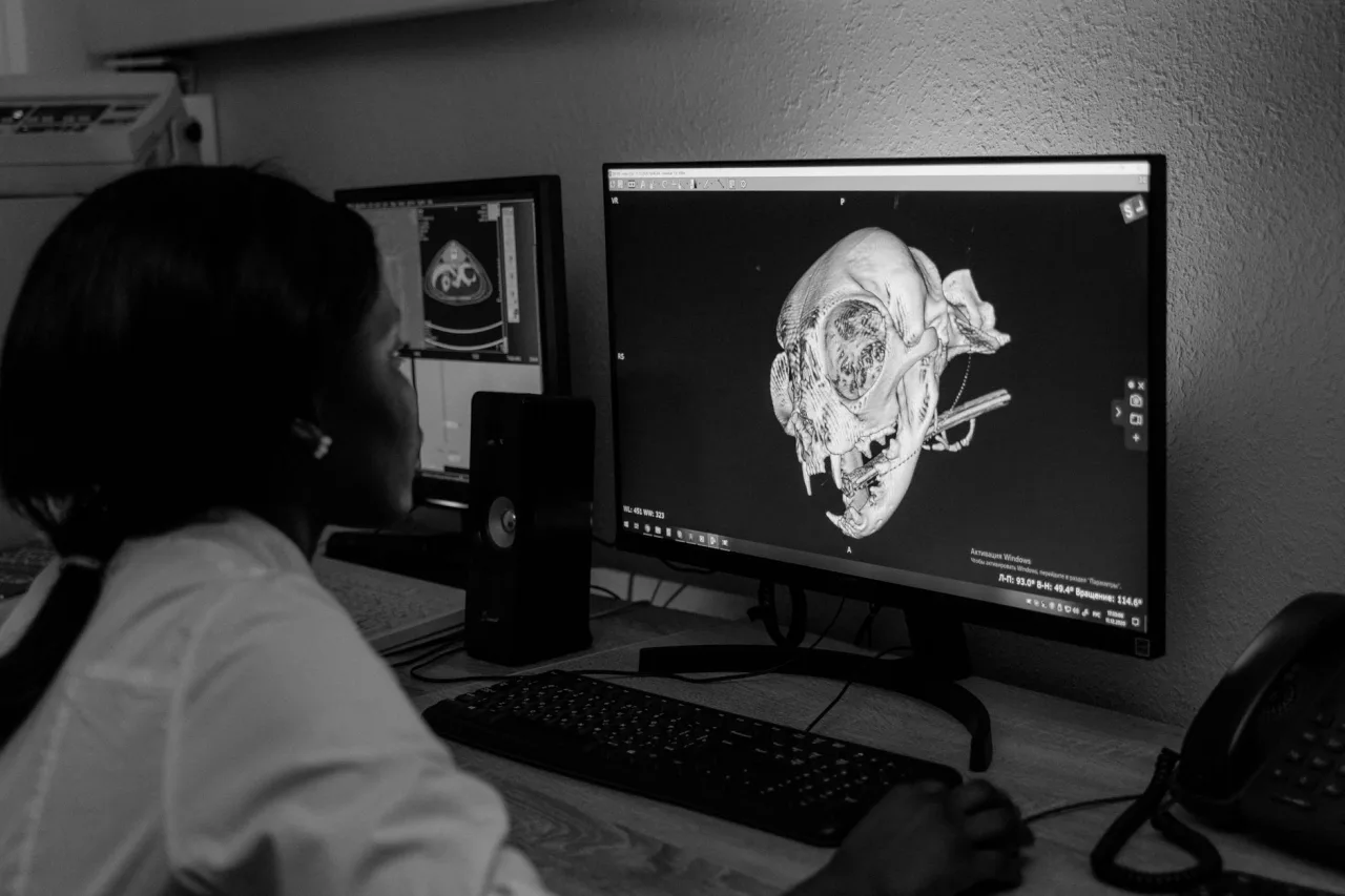
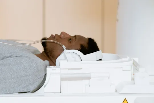
Most visits share a shape. You check in and confirm your identity and the exact test. You may change into a gown and remove jewelry, glasses or anything metal, which matters most for MRI. The technologist positions you, runs the scan, and may take several views or sequences. Timing ranges widely: a plain X-ray takes a few minutes, ultrasound and CT often run 15 to 45 minutes, and MRI can take 30 to 60 minutes or longer because it captures multiple sequences.
If enclosed spaces make you anxious, say so when you book, because wide-bore and open MRI options and other accommodations exist. If you are or might be pregnant, tell the team before any scan, because it can change which test is used.
Preparation is not one-size-fits-all
- Some abdominal ultrasounds and CT scans require fasting for several hours.
- A pelvic ultrasound may need a full bladder, so you drink water and hold it.
- Contrast dye (by mouth, injection or both) is used for some CT and MRI exams to make vessels or organs stand out.
- Tell staff about pregnancy, kidney problems, allergies, diabetes medicine, or any implanted metal or device before the scan.
Is imaging safe?
Safety depends on the method. Ultrasound uses sound waves and MRI uses a magnetic field and radio waves, so neither involves ionizing radiation, which is one reason ultrasound is favored in pregnancy. X-ray, CT and mammography do use ionizing radiation, but in low, carefully chosen doses. RadiologyInfo.org notes that radiologists and technologists are trained to use the smallest amount of radiation necessary to obtain the images, and that when an exam is needed to assess your health, the benefits greatly outweigh any risk. Modern equipment has further reduced exposure while keeping image quality high.
The practical takeaway is that the small risk of a well-chosen scan is almost always outweighed by the value of an accurate diagnosis. If avoiding radiation matters for your situation, it is reasonable to ask your doctor whether a radiation-free alternative such as ultrasound or MRI would answer the same question.
What contrast dye does
Contrast agents temporarily improve the visibility of specific structures. Iodine-based contrast is common in CT, gadolinium-based contrast is used in some MRI exams, and barium is sometimes swallowed to image the digestive tract. Reactions are uncommon and usually mild, but the team will ask about allergies and kidney function first, because the kidneys clear many of these agents. Not every scan uses contrast; your doctor decides based on what they need to see.
Making sense of your report (and its limits)
A typical radiology report describes the technique used, the findings, and an impression that summarizes what the findings suggest. You may see words like "unremarkable," which means normal, "no acute findings," or a description of something that needs follow-up. Reading these lines out of context is where worry usually starts.
Two honest cautions. First, imaging findings often need to be interpreted alongside your symptoms, physical examination and other test results; an image rarely tells the whole story alone. Second, incidental findings, meaning something unexpected and usually harmless, are common and can trigger further tests. The person best placed to explain your report, and what to do next, is the doctor who ordered it.
General information, not medical advice
- This article is educational. It does not diagnose any condition or interpret your specific results.
- Only your referring clinician or radiologist can tell you what your images mean for you.
- If you have severe or sudden symptoms, seek urgent medical care rather than waiting for a scheduled scan.
Choosing where to have your scan
You often have a choice of facility. Sensible things to weigh include whether the center is accredited, whether it has the exact equipment your doctor requested, how quickly it can send the report to your clinician, and the total cost including any contrast or facility fees. Hospital radiology departments and dedicated outpatient imaging centers both do excellent work; the right fit depends on your test, your insurance and your convenience. Our directory of imaging centers explains what to look for when you compare options.
On cost, be cautious with any exact figure you see online. Prices vary widely by country, provider and insurance, and the same scan can carry very different prices even within one city. Ask for the total expected cost, whether pre-authorization is needed, and what your insurer will cover, then confirm those numbers with both the clinic and your insurer before the appointment.
Questions worth asking your doctor
- Why is this specific scan the best choice for my symptoms?
- Is there a radiation-free alternative that would answer the same question?
- Do I need contrast, and does anything about my health affect that?
- How and when will I get my results, and who will explain them to me?
Frequently asked questions
What is the difference between imaging and radiology?
The terms overlap. Imaging, or diagnostic imaging, refers to the tests that create pictures of the inside of the body, while radiology is the medical specialty devoted to performing and interpreting those tests. A radiologist is the doctor who reads the images and writes the report your own clinician then discusses with you.
Which imaging tests use radiation and which do not?
X-ray, CT and mammography use small, controlled doses of ionizing radiation. Ultrasound and MRI do not use ionizing radiation: ultrasound uses sound waves and MRI uses a magnetic field and radio waves. If avoiding radiation matters for your situation, ask your doctor whether one of these alternatives would answer the same question.
How should I prepare for imaging?
It depends entirely on the test. Some scans require fasting, a full bladder, or pausing certain medicines, while others need no preparation at all. The clinic will give you specific instructions when you book, and following them closely helps you avoid a repeat visit.
How long does it take to get imaging results?
Timing varies by facility and by how urgent the test is. Some results are available within hours, while routine studies may take several days to be reported and sent to your referring doctor. That doctor will contact you to discuss the findings and any next steps.
Can imaging tell me exactly what is wrong?
Not always on its own. Imaging is a powerful clue, but findings usually need to be interpreted alongside your symptoms, examination and other tests. Your clinician combines all of that information to reach a diagnosis, which is why you should discuss your report with them rather than acting on it alone.
Is it safe to have imaging during pregnancy?
Ultrasound and MRI, which use no ionizing radiation, are generally preferred during pregnancy, while tests that use radiation are used only when the benefit clearly justifies it. Always tell the team if you are or might be pregnant so they can choose the safest appropriate test and answer your questions before the scan.
Authoritative sources used for this article include MedlinePlus on diagnostic imaging and RadiologyInfo.org on radiation dose safety. Always confirm what applies to you with your own healthcare provider.
More guides on Ultrasound & Radiology (General)
Have a question or a topic to suggest?
We read every message. Tell us what you would like to see next.
You may also like
CT Imaging Near Me: Compare Clinics
Holding a CT referral and choosing between centers? Use a simple scorecard to compare accreditation, reporting, price, and turnaround instead of picking the first result.
DEXA Scan Near Me: Choose a Clinic
A DEXA scan is one test where the clinic you pick has a lasting effect, because results are compared over years. Learn how to choose a good bone density center.

Ultrasound Locations Near Me
Hospitals, imaging centers, and keepsake studios all advertise ultrasound. Learn to tell a proper diagnostic location from a novelty one before you book.