X-Ray Scan
An X-ray scan is one of the fastest and most common medical imaging tests, often done in minutes to check bones, the chest and more.

An X-ray scan is one of the fastest and most common medical imaging tests, often done in minutes to check bones, the chest and more. This guide explains what an X-ray exam involves, why doctors order it, how much radiation it actually uses, and how the results reach you, so you can walk in prepared and calm.
The short version
- An X-ray scan sends a small, controlled amount of radiation through the body to create images of bones and some organs.
- It is quick, painless and widely used for fractures, chest problems, dental checks and more.
- The radiation dose is low. A chest X-ray is roughly the natural background radiation you get over about 10 days.
- A radiologist reads the images and reports to your doctor, who explains what they mean for you.
What an X-ray scan is
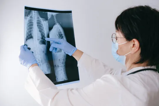
An X-ray uses a form of energy that passes through the body to create a picture. MedlinePlus explains that "X-rays send a small amount of radiation through your body to create pictures of bones and organs." Dense tissue such as bone absorbs more and appears white, soft tissue appears grey, and air-filled spaces such as the lungs appear dark. That contrast is what lets a radiologist spot a fracture, a shadow on the lung or a swallowed object.
The plain X-ray is the base of a larger family of tests that all use the same physics. If you want the wider picture of how these technologies relate, read our companion guide to X-ray imaging, and for the overall field see what radiology covers.
Why doctors order an X-ray scan

X-rays answer many everyday clinical questions quickly. The NHS lists common uses including checking bones for fractures, looking at teeth for decay, spotting problems in the digestive system and checking whether a child has swallowed something dangerous. MedlinePlus adds chest imaging to detect pneumonia and other lung conditions, and notes that mammograms are a specialised X-ray used for breast cancer screening.
- Bones and joints: suspected fractures, dislocations and arthritis.
- Chest: the lungs, heart size and signs of infection such as pneumonia.
- Abdomen: some blockages and swallowed objects.
- Dental and screening: tooth decay and mammography.
For specific studies, we have detailed guides such as the chest X-ray for pneumonia and the hip X-ray.
How much radiation is involved
This is the question most people worry about, and the reassuring answer is that a plain X-ray dose is low. RadiologyInfo measures radiation in units called millisieverts (mSv) and gives a single chest X-ray as about 0.1 mSv, roughly the same as 10 days of natural background radiation. For context, the average person receives about 3 mSv per year from natural sources such as cosmic rays and radon. MedlinePlus makes the same comparison, describing a chest X-ray as similar to about 10 days of everyday background exposure.
Facilities keep doses low using the ALARA principle, which RadiologyInfo defines as "As Low As Reasonably Achievable." The NHS describes X-rays as usually very safe with rare serious complications, while acknowledging that frequent exposure carries a very small long-term risk. Doctors weigh that small risk against the clear benefit of an accurate diagnosis before ordering the test.
Always mention pregnancy. MedlinePlus advises telling your healthcare provider if you are or might be pregnant before an X-ray. The team can then decide whether to adjust, shield or use a different test. When in doubt, say something before the scan.
How to prepare

Most plain X-rays need little preparation. You may be asked to remove jewellery, glasses and any clothing with metal near the area being imaged, since metal shows up and can obscure the picture. You might change into a gown. Some studies that use a contrast agent, such as certain digestive or urinary exams, have specific eating or drinking instructions, so follow whatever your clinic tells you.
What happens during the scan
A radiographer positions the body part against or near the detector and may ask you to stand, sit or lie down. You hold still, and for a chest X-ray you may be asked to take a breath and hold it. The exposure itself lasts a fraction of a second, and you feel nothing. Several views from different angles may be taken. The whole visit is usually over in a few minutes. You can go about your day immediately afterward, and there is no radiation left in your body.
What the results mean
The images are read by a radiologist, who writes a report for the doctor who ordered the scan. That doctor then discusses the findings with you alongside your symptoms and history. An X-ray shows a great deal about bone and the chest, but it does not show everything, and a normal X-ray does not always rule out a problem. If a finding needs a closer look, your doctor may order an ultrasound, a CT or an MRI. Our comparison of CT and MRI explains how those tests differ. Do not try to interpret the images yourself.
Costs vary by country, provider and insurance. A plain X-ray is usually one of the cheaper imaging tests, but always confirm the price with the clinic and your insurer, and if you need to locate a provider you can compare an X-ray facility near you.
This is general information, not medical advice. This article explains X-ray scans in general terms. It cannot tell you whether you need one or what your images mean. Please talk with your referring clinician or a radiologist about your own situation.
Frequently asked questions
Is an X-ray scan safe?
For a single plain X-ray the dose is low, about 0.1 mSv for a chest film, which RadiologyInfo compares to roughly 10 days of natural background radiation. The NHS describes X-rays as usually very safe with rare serious complications. Frequent exposure carries a very small long-term risk, so doctors order X-rays only when the benefit outweighs it.
How long does an X-ray scan take?
The exposure lasts a fraction of a second, and the whole visit is usually just a few minutes, including positioning and taking a few views. Studies that use contrast can take longer. You can return to normal activity straight away.
Does an X-ray scan hurt?
No. The scan itself is painless and you feel nothing during the exposure. Any discomfort usually comes from holding a position with an injured body part, and the radiographer will help you get as comfortable as possible.
Should I tell anyone if I might be pregnant?
Yes. MedlinePlus advises telling your healthcare provider if you are or might be pregnant before an X-ray. The team can then decide whether to adjust the exam, use shielding or choose a different test.
What happens after the X-ray?
A radiologist reviews the images and sends a report to the doctor who ordered the scan, who then explains the findings in the context of your symptoms. There is no radiation left in your body, so you can carry on with your day immediately.
More guides on MRI, CT & X-Ray
Have a question or a topic to suggest?
We read every message. Tell us what you would like to see next.
You may also like
MRI Tech
When people search for "MRI tech," they usually mean one of two things: the technology that makes a magnetic resonance imaging scan possible, or the technologist who runs the machine.

MRI Testing
MRI testing can feel mysterious the first time: a referral appears, an appointment gets booked, and you are told to lie still inside a loud tunnel.
Stand Up MRI
A stand up MRI, sometimes called an upright, positional or weight-bearing MRI, lets you be scanned sitting, standing or bending rather than only lying flat.