Perfusion Imaging
Perfusion imaging is a family of scans that measure blood flow through living tissue rather than just showing its shape.
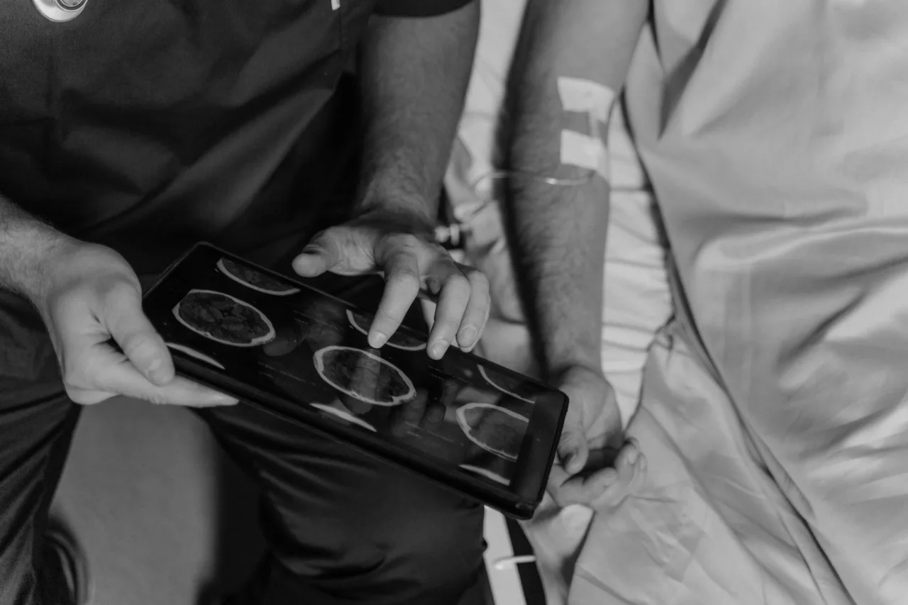
Perfusion imaging is a family of scans that measure blood flow through living tissue rather than just showing its shape. By tracking how blood moves into and out of an organ, these tests help doctors judge whether tissue is getting enough supply, most often in the brain and the heart. This guide explains what perfusion imaging is, how it is done, and how the results are used, though only your own clinician can interpret what a scan means for you.
The short version
- Perfusion means the passage of blood through the small vessels of a tissue, and perfusion imaging measures that flow.
- It can be done with CT, MRI, or nuclear medicine, so the radiation and contrast involved depend on which method is used.
- Common uses include assessing stroke, checking blood supply to the heart muscle, and studying some tumours.
- A radiologist reads perfusion maps together with other images and your history, so the colours and numbers are only part of the answer.
What perfusion imaging actually is
Perfusion is the passage of blood through the small vessels that feed a tissue or organ. According to the overview on Wikipedia, perfusion scanning observes, records, and measures that flow, producing maps and graphs of values such as blood flow rate and blood volume over time. Instead of a still picture of anatomy, perfusion imaging shows how well blood is actually reaching the tissue.
This makes it a functional test, one that measures activity rather than only structure. It sits within the wider field of medical imaging, and it is usually added to a more conventional scan such as a CT or MRI of the same area, interpreted as part of the broader radiology workflow.
The main ways perfusion is measured
There is no single perfusion machine. The technique can be run on several different systems, and the choice affects speed, availability, and whether radiation or contrast is involved. The Wikipedia overview describes the main approaches:
- CT perfusion (CTP): an iodine-based contrast dye is injected and a CT scanner takes rapid, repeated images to follow the dye through the tissue. It is fast and widely available, and it uses X-rays.
- MR perfusion (MRP): uses an MRI scanner, either with a gadolinium contrast agent or with a contrast-free method that magnetically labels the blood itself. It uses no ionising radiation.
- Nuclear medicine perfusion: a radioactive tracer is taken up in proportion to blood flow, and a gamma camera or PET scanner records where it goes. This is the basis of many heart and lung studies.
Because the methods differ, your clinician and the imaging team choose the one that best answers the clinical question. Our overviews of the CT scan and the MRI scan explain the two most common platforms in more detail.
Why doctors order perfusion imaging
Perfusion imaging is valuable whenever the key question is whether tissue is receiving enough blood. Established uses include:
- Stroke: the American College of Radiology resource RadiologyInfo notes that CT perfusion and MR perfusion can show blood flow to help physicians choose the best treatment in acute stroke.
- Heart: myocardial perfusion studies look at blood supply to the heart muscle, often comparing rest and stress, which complements a structural test like a heart ultrasound.
- Tumours: perfusion can help characterise abnormal blood supply in some masses and monitor how they respond over time.
- Lungs: a ventilation and perfusion study compares airflow with blood flow, for example when a clot in the lung is suspected.
In every case the scan is descriptive. It gives the team a measurement of flow that feeds into a decision, alongside your symptoms, examination, and other tests.
How a perfusion scan is performed
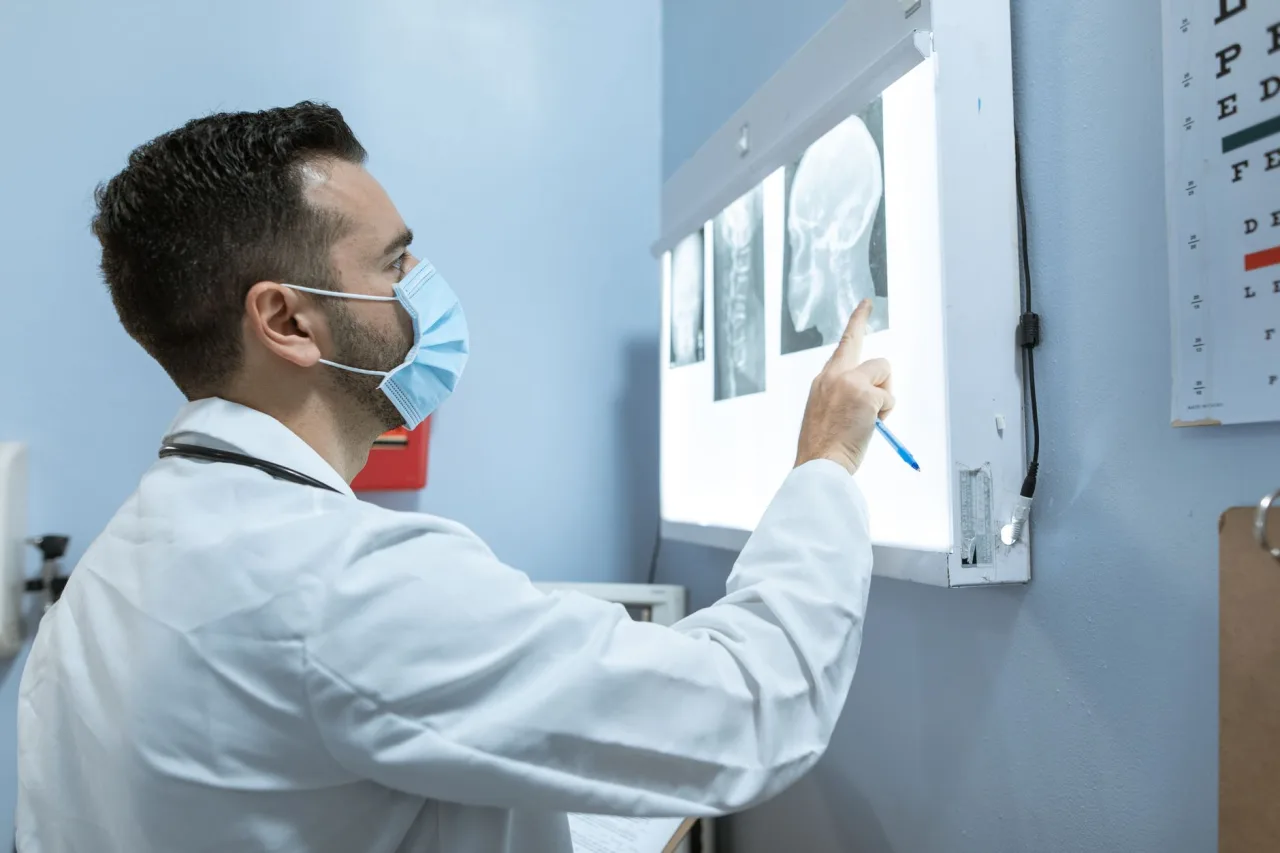
The exact steps depend on the method, but a contrast-based CT or MR perfusion study of the brain generally follows a pattern like this:
- A cannula is placed in a vein so contrast can be injected quickly during the scan.
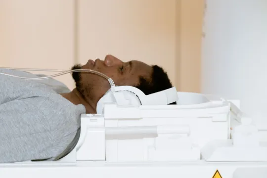
- You lie still on the scanner table, and the area of interest is positioned in the machine.
- The contrast is injected, often by a pump, and the scanner captures a rapid series of images as it passes through the tissue.
- Software converts the changing signal into maps of blood flow, blood volume, and timing.
- A radiologist reviews these maps together with the standard images from the same visit.
Good to know: A contrast-free MR perfusion method called arterial spin labelling uses the blood itself as a natural tracer, so it can avoid an injection. Whether it is suitable depends on the clinical question and the scanner.
Is perfusion imaging safe?
Safety depends entirely on which method is used. CT perfusion uses X-rays and an iodine-based contrast dye, so it carries a small radiation dose and the usual precautions around contrast. MR perfusion uses no ionising radiation, and if it uses gadolinium contrast the team will check your kidney function and allergy history first, as the NHS describes for MRI in general. Nuclear perfusion studies use a small radioactive tracer. Tell the team if you are pregnant or breastfeeding, have kidney problems, or have had reactions to contrast, so they can choose the safest option.
What perfusion results mean
Perfusion imaging produces colour maps and numbers that represent flow, volume, and timing in the tissue. These are powerful, but they are only meaningful when read by a radiologist alongside the anatomical images and your clinical situation. A pattern that looks abnormal on a map may reflect the disease in question, a technical artefact, or normal variation, and telling these apart requires expertise. A perfusion study does not provide a diagnosis on its own, and a normal result does not exclude every condition.
This is why interpretation belongs to a qualified clinician. Trying to read your own maps or report can cause unnecessary worry or false reassurance. If you have had a perfusion scan, bring your questions to the doctor who ordered it.
Please note: This article is general educational information, not medical advice, and it cannot tell you what your own scan means. For anything about your symptoms, your scan, or your results, speak with your doctor, referring clinician, or a radiologist.
How perfusion fits with other scans
Perfusion imaging almost always works alongside other studies rather than replacing them. In a stroke assessment, for example, a plain CT or MRI shows the anatomy, an angiogram shows the larger blood vessels, and perfusion shows the flow within the tissue. Similarly, a brain MRI may combine perfusion with sequences like susceptibility weighted imaging, which is tuned to tiny bleeds. Reading these together gives a fuller picture than any one method alone. If you need to arrange a scan, our guide to finding a radiology clinic near you explains what to look for.
Frequently asked questions
What is the difference between a normal scan and a perfusion scan?
A normal CT or MRI mostly shows the shape and structure of tissue. A perfusion scan measures blood flow through that tissue over time, producing maps of how well blood is reaching it. The two are often done together and read side by side.
Does perfusion imaging use radiation?
It depends on the method. CT perfusion and nuclear perfusion studies involve some radiation, while MR perfusion uses none because MRI relies on magnets and radio waves. Your clinician chooses the method based on the question and your circumstances.
Will I need a contrast injection?
Often yes. CT perfusion uses an iodine-based dye, and MR perfusion frequently uses a gadolinium agent, though a contrast-free MRI method exists. The imaging team will explain what is planned and check for allergies and kidney issues beforehand.
What is perfusion imaging most commonly used for?
Common uses include assessing acute stroke, checking blood supply to the heart muscle, studying some tumours, and comparing airflow with blood flow in the lungs. A radiologist interprets the results in the context of your history and other tests.
Can perfusion imaging diagnose my condition by itself?
No. It provides measurements of blood flow that support a diagnosis, but it is read alongside other images and your clinical picture. Only a qualified clinician can put the findings together and tell you what they mean for you.
More guides on Ultrasound & Radiology (General)
Have a question or a topic to suggest?
We read every message. Tell us what you would like to see next.
You may also like
CT Imaging Near Me: Compare Clinics
Holding a CT referral and choosing between centers? Use a simple scorecard to compare accreditation, reporting, price, and turnaround instead of picking the first result.
DEXA Scan Near Me: Choose a Clinic
A DEXA scan is one test where the clinic you pick has a lasting effect, because results are compared over years. Learn how to choose a good bone density center.

Ultrasound Locations Near Me
Hospitals, imaging centers, and keepsake studios all advertise ultrasound. Learn to tell a proper diagnostic location from a novelty one before you book.