Muga Scan
A MUGA scan is a nuclear imaging test that shows how strongly your heart pumps blood with each beat.
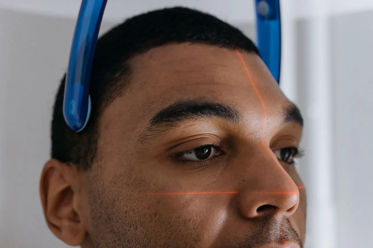
A MUGA scan is a nuclear imaging test that shows how strongly your heart pumps blood with each beat. Its main job is to measure the ejection fraction, a percentage that reflects the pumping strength of the heart's main chamber. This guide explains why the test is ordered, how it is performed, and how to think about the results, though only your own clinician can interpret what a scan means for you.
The short version
- MUGA stands for multigated acquisition. It is a nuclear medicine test, also called radionuclide angiography or radionuclide ventriculography.
- A small amount of radioactive tracer is attached to your red blood cells so a special camera can watch blood move through the heart's chambers.
- The headline number is the ejection fraction, and a resting value of roughly 50 to 75 percent is often described as a typical range.
- A cardiologist reads the scan alongside your history and other tests, so never judge your own result from a single number.
What a MUGA scan actually is
MUGA is short for multigated acquisition, and the same test is also called radionuclide angiography or radionuclide ventriculography. The American Heart Association describes it as a type of nuclear imaging test that shows how well the heart pumps. Unlike a plain picture of the heart's shape, a MUGA scan tracks moving blood, which lets the equipment calculate how much of the blood in the main pumping chamber is pushed out with each beat.
The test belongs to the branch of medical imaging called nuclear medicine, where a tiny quantity of a radioactive substance called a tracer is introduced into the body and a gamma camera detects where it goes. If you want the wider picture of how the different scan families fit together, our overview of medical imaging sets the scene, and the general radiology guide explains who reads these studies.
Why doctors order a MUGA scan
A clinician usually requests a MUGA scan when they need a reliable measure of the heart's pumping strength. The Cleveland Clinic notes that it is often used to check for heart failure and, importantly, to watch the heart during cancer treatment, because some chemotherapy drugs can weaken the heart muscle over time. Common reasons include:
- Assessing symptoms such as breathlessness, fatigue, chest discomfort, or swelling that may point to reduced pumping strength.
- Measuring the ejection fraction before, during, and after chemotherapy so the care team can catch early signs of heart strain.
- Evaluating known or suspected coronary artery disease, valve disease, or cardiomyopathy.
- Following up on an abnormal result from another test, such as an electrocardiogram.
A MUGA scan is descriptive rather than a treatment. It gives the team a precise measurement so they can decide whether medication, a change in cancer therapy, or further tests are needed. A related first-line structural test is the heart ultrasound, which also reports an ejection fraction using sound waves instead of a tracer.
How the scan works, step by step
Knowing the sequence can ease any nerves. A standard resting MUGA scan generally follows these stages.
- Small sticky electrodes are placed on your chest to record your heartbeat. The camera uses this signal to synchronise, or gate, its pictures to each phase of the beat.
- A small amount of technetium-99m tracer is injected into a vein. In many protocols the tracer is prepared so that it attaches to your own red blood cells.
- After a short wait for the tracer to spread evenly through the bloodstream, you lie still under a gamma camera.
- The camera records images from several angles as your heart beats, often front, angled, and side views, so the software can outline the pumping chamber.
- A computer combines many heartbeats into a representative cycle and calculates the ejection fraction.
The scan itself is painless apart from the needle for the injection. The Cleveland Clinic notes that the appointment commonly takes about one to three hours, partly because of the wait for the tracer to circulate.
Good to know: The tracer is a very small dose and is not a contrast dye of the kind used in some CT studies. It is designed to leave the body naturally, largely through the kidneys, over roughly a day.
Resting and exercise MUGA
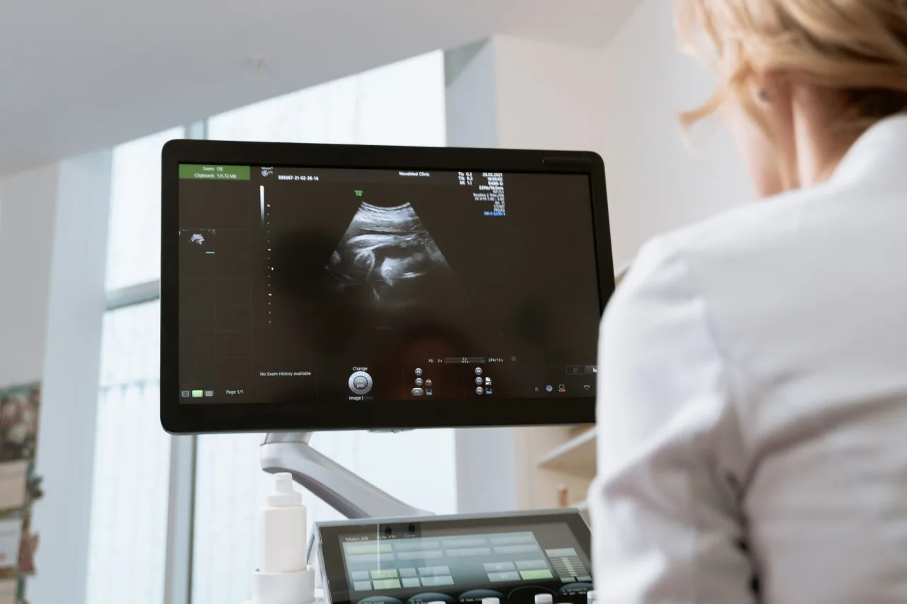
There are two broad versions of the test. A resting MUGA measures pumping strength while you lie quietly. An exercise, or stress, MUGA compares the heart at rest with the heart while it works harder, sometimes using a treadmill or a stationary cycle between imaging runs. Comparing the two states can reveal problems that only appear under exertion, such as areas of muscle that do not respond normally when demand rises. Your clinician decides which version answers the question they have, based on your symptoms and history.
How to prepare
Preparation is usually simple, but instructions vary by clinic and country, so always follow the exact guidance from the department that booked your test. General points often include:
- You may be asked to avoid caffeine and certain heart medicines for a set period before a stress study, because they can affect the heart's response. Never stop a prescribed medicine without being told to.
- Wear comfortable, two-piece clothing, and shoes suitable for walking if an exercise portion is planned.
- Tell the team if you are pregnant or breastfeeding, or think you might be, before any tracer is given.
- Mention all medicines, allergies, and recent scans, since other nuclear studies can occasionally interfere.
Is a MUGA scan safe?
A MUGA scan does involve a small amount of radiation, because it uses a radioactive tracer and is a nuclear medicine test. Both the American Heart Association and the Cleveland Clinic describe the tracer as safe for most people, with the dose leaving the body within about a day. Serious reactions are uncommon. The main caution is pregnancy and breastfeeding, where the test is generally avoided or delayed unless the benefit clearly outweighs the risk, which is a decision for your clinician.
Because it uses a tracer rather than magnets or sound waves, a MUGA scan sits in a different safety category from a radiation-free MRI scan or a heart ultrasound. A CT scan also uses radiation but works in a completely different way. Your care team weighs these trade-offs when choosing a test.
What ejection fraction results mean
The central result of a MUGA scan is the left ventricular ejection fraction, the percentage of blood pushed out of the main chamber with each beat. The Cleveland Clinic describes a value of roughly 50 to 75 percent as a commonly quoted typical range, with lower values suggesting the heart is pumping less blood than expected. A result outside a reference range is not an automatic diagnosis, though, and a single number always needs the context of your symptoms, examination, and other investigations.
This is why interpretation belongs to a qualified clinician. Trying to read your own report can cause needless worry or false reassurance. If you have been given a figure, bring your questions to the cardiologist or referring clinician who ordered the test.
Please note: This article is general educational information, not medical advice, and it cannot tell you what your own scan means. For anything about your heart, symptoms, medicines, or results, speak with your doctor, referring clinician, or a cardiologist.
How MUGA compares with other heart tests
A MUGA scan is one of several ways to measure heart function, and each has strengths:
- Heart ultrasound (echocardiogram): uses sound waves, no radiation, widely available, and also reports an ejection fraction while showing valves and chamber sizes.
- Cardiac MRI: gives very detailed tissue images using magnets and no radiation, often reserved for specific questions.
- MUGA scan: historically valued for a highly reproducible ejection fraction, which is useful for tracking small changes during chemotherapy.
Because these tests answer overlapping questions in different ways, the choice depends on what needs to be measured, how precisely, and how often. If you are trying to arrange a study, our guide to finding a radiology clinic near you explains what to look for.
Frequently asked questions
Does a MUGA scan hurt?
The scan itself is painless. The only discomfort is usually the small needle used to inject the tracer into a vein. Lying still under the camera can feel long, but it does not hurt.
How much radiation does a MUGA scan involve?
A MUGA scan uses a small amount of a radioactive tracer, so there is some radiation exposure, unlike an ultrasound or MRI. The American Heart Association and Cleveland Clinic describe the tracer as safe for most people, leaving the body within about a day. Your clinician balances this small exposure against the value of the information.
How long does the test take?
The appointment often runs about one to three hours according to the Cleveland Clinic, mostly because of the wait for the tracer to circulate before images are taken. The imaging itself is a smaller part of that time.
What is a normal ejection fraction on a MUGA scan?
A resting left ventricular ejection fraction of roughly 50 to 75 percent is a commonly quoted typical range, with lower values suggesting weaker pumping. A number on its own is not a diagnosis. Only your clinician can say what a specific value means for you.
Can I have a MUGA scan if I am pregnant?
Because the test uses a radioactive tracer, it is generally avoided during pregnancy and breastfeeding unless the benefit clearly outweighs the risk. Always tell the team if you are or might be pregnant so they can choose the safest approach.
More guides on Ultrasound & Radiology (General)
Have a question or a topic to suggest?
We read every message. Tell us what you would like to see next.
You may also like
CT Imaging Near Me: Compare Clinics
Holding a CT referral and choosing between centers? Use a simple scorecard to compare accreditation, reporting, price, and turnaround instead of picking the first result.
DEXA Scan Near Me: Choose a Clinic
A DEXA scan is one test where the clinic you pick has a lasting effect, because results are compared over years. Learn how to choose a good bone density center.

Ultrasound Locations Near Me
Hospitals, imaging centers, and keepsake studios all advertise ultrasound. Learn to tell a proper diagnostic location from a novelty one before you book.