Color Doppler Ultrasound
A color Doppler ultrasound is a standard ultrasound with an added trick: it shows blood flowing through your vessels and organs in real time
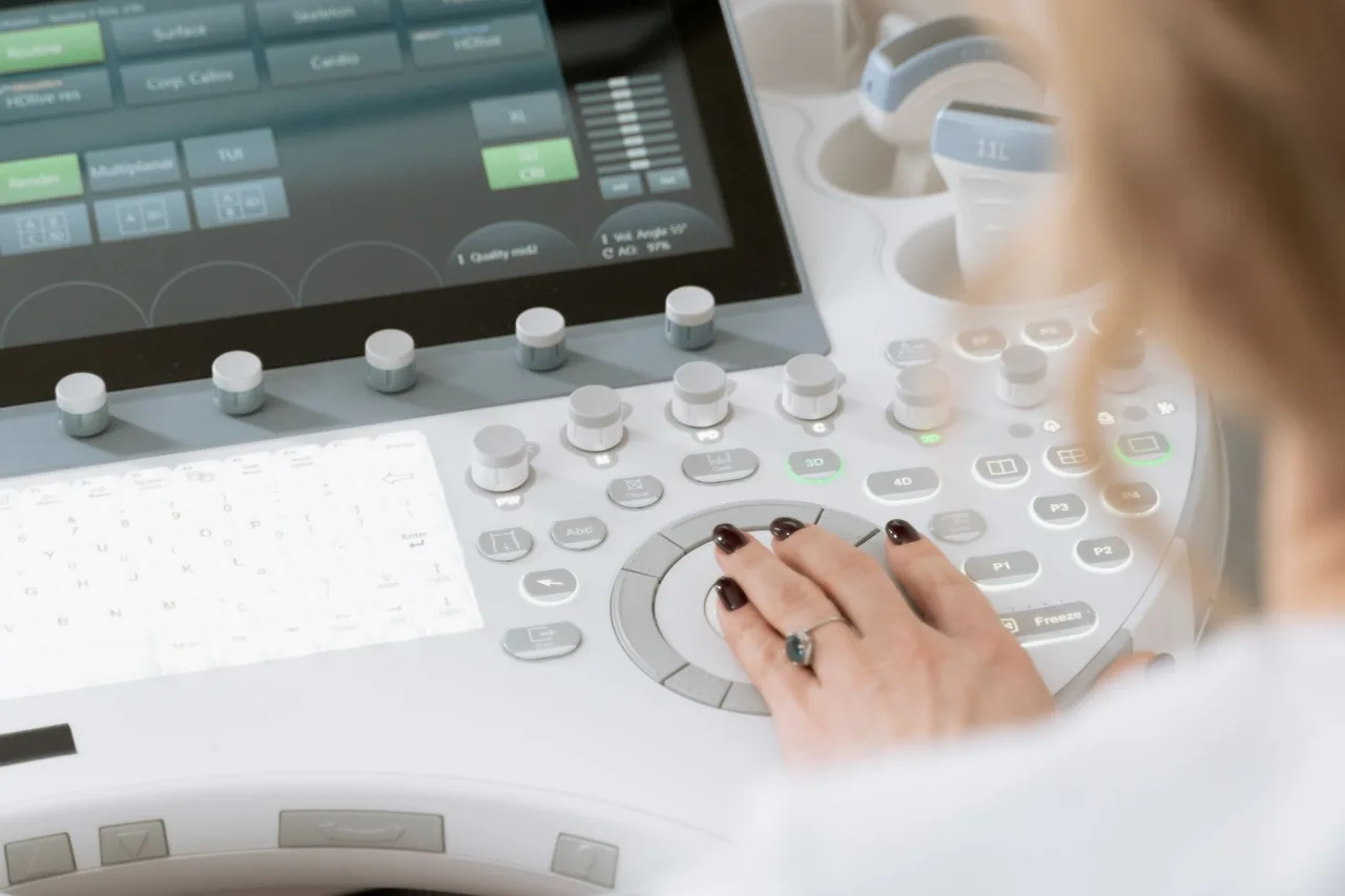
A color Doppler ultrasound is a standard ultrasound with an added trick: it shows blood flowing through your vessels and organs in real time, painted in color on the screen. It uses the same safe sound waves as any other ultrasound, with no radiation and no needles. This guide explains what color Doppler is, why doctors order it, how the scan is done, and how to make sense of the report.
The short version
- Color Doppler is an ultrasound technique that measures moving blood and displays its speed and direction as color over the normal grey scale image.
- It is used to check for blocked or narrowed arteries, clots in veins, blood flow to organs and the fetus, and the workings of the heart.
- The scan is painless, radiation free, and done exactly like a regular ultrasound, usually taking thirty to sixty minutes.
- The colors and numbers are technical measurements. Only a radiologist and your doctor can interpret what they mean for you.
What color Doppler ultrasound is
Ordinary ultrasound sends sound waves into the body and turns the returning echoes into a grey scale picture of tissue. Doppler ultrasound goes a step further by measuring how the frequency of those echoes shifts when they bounce off moving red blood cells. That shift reveals how fast blood is moving and in which direction. A color Doppler scan converts these measurements into color, so a physician can see circulation through vessels at a glance, overlaid on the normal image.
According to RadiologyInfo, there are a few related forms of the technique. Color Doppler maps flow as color, power Doppler is a more sensitive version that detects very slow or faint flow but cannot show direction, and spectral Doppler plots flow as a graph or a sound over time. Many scans combine these. For the basics of the underlying technology, see our explainer on what an ultrasound is and how it works.
Why a doctor orders one

Color Doppler is added whenever the question is not just what an organ looks like, but how blood is moving through it. Common reasons include:
- Checking the leg or arm veins for a deep vein thrombosis, a blood clot.
- Assessing the carotid arteries in the neck for narrowing that can raise stroke risk.
- Looking for narrowed or blocked arteries causing pain or poor circulation.
- Evaluating blood flow to organs such as the liver, kidneys or a transplanted organ.
- Monitoring blood flow to the placenta and fetus during pregnancy.
- Studying the heart and its valves, where it is part of an echocardiogram.
When the heart is the focus, Doppler is central to a heart ultrasound. Doppler is also frequently added during a pelvic ultrasound to assess blood supply to the ovaries, uterus or, in men, the testicles.
How to prepare
Preparation depends on the part of the body being examined, so the most important step is to follow your clinic's instructions. As a general guide:
- For most limb, neck or vascular scans, no special preparation is needed, and you can eat and take your medicines as usual.
- For a scan of the abdominal blood vessels or organs, you may be asked to fast for several hours to reduce bowel gas.
- Wear loose, comfortable clothing so the area can be uncovered easily.
- You may be asked to avoid caffeine or nicotine before some vascular studies, as these can affect blood vessels.
Tip: Bring the details of any previous Doppler scan or vascular procedure. Comparing new measurements with earlier ones helps the radiologist judge whether flow has genuinely changed over time.
What happens during the scan

A color Doppler scan feels exactly like a regular ultrasound. You lie on a couch, the sonographer applies warm gel to the skin, and moves a probe over the area while watching the screen. When Doppler is switched on you may hear a rhythmic, pulsing or whooshing sound, which is the blood flow turned into audio. This is completely normal and can help the sonographer find and follow a vessel.
The sonographer may press a little more firmly over a vein to see whether it compresses, or ask you to breathe in a certain way, change position, or hold still while a measurement is taken. A full study can take longer than a plain ultrasound, often thirty to sixty minutes, because each vessel is examined carefully. The test is painless, and when it is finished the gel is wiped off and you can return to your usual activities.
Understanding your results
A radiologist reviews the images, the color maps and the flow measurements, then writes a report for the doctor who referred you. You will usually not get results during the appointment. The report is technical: it may describe normal flow, a narrowing expressed as a percentage, an increased flow velocity, a clot that stops a vein compressing, or reduced flow to an organ.
These are measurements and observations, not a diagnosis on their own. Their meaning depends on your symptoms, your history and sometimes other tests. A finding that is important in one person may be minor in another, which is exactly why the interpretation belongs with your clinician rather than with a number on a screen.
Important: This article gives general educational information and is not medical advice. It cannot tell you what your own Doppler results mean. Please discuss your report, symptoms and any next steps with your referring doctor or a radiologist.
Benefits, limits and safety
Color Doppler is safe, painless and uses no ionising radiation, so it can be repeated freely and is suitable in pregnancy. It is widely available, gives real time information about circulation, and often avoids the need for more invasive tests such as angiography with dye.
Its limits come from physics and technique. Doppler is angle dependent, so the probe must be aligned well with the vessel for accurate readings, and results rely heavily on the skill of the operator. Sound waves are also blocked by gas and bone and weaken with depth, so bowel gas, a large body habitus or deep vessels can reduce accuracy. When Doppler cannot fully answer the question, your doctor may add a CT scan or another test.
How to find a good clinic
Because Doppler is so operator dependent, the quality of the clinic matters even more than usual. Look for accreditation, scans performed by qualified sonographers with vascular experience, and reports written by a radiologist. Ask how quickly your report will reach your doctor and whether images are stored for future comparison. Our guide on how to find sonography services near you covers what to check before you book.
Prices vary by country, provider and whether you pay privately or use insurance, so confirm the fee, what it includes and your coverage in advance. A scan that includes a full radiologist interpretation generally offers better value than a cheaper scan without one.
Frequently asked questions
What is the difference between an ultrasound and a color Doppler?
A standard ultrasound shows what tissue and organs look like in grey scale. A color Doppler adds a measurement of moving blood and paints its speed and direction in color over the same image. Doppler is not a separate machine, it is a mode used on the same ultrasound scanner.
Is a color Doppler ultrasound safe?
Yes. It uses the same sound waves as any ultrasound, with no ionising radiation and no injections for a standard scan. It has no known harmful effects and can be repeated as needed, including during pregnancy when performed for a genuine clinical reason.
Why do I hear a whooshing sound during the scan?
That sound is your blood flow converted into audio by the Doppler system. It is completely normal and helps the sonographer locate and follow a vessel. The pattern and pitch of the sound also give the sonographer information about how the blood is moving.
How long does a color Doppler scan take?
A full Doppler study often takes about thirty to sixty minutes, longer than a plain ultrasound, because each vessel is examined and measured carefully. The exact time depends on the area being studied and how many vessels need to be checked.
What can a color Doppler ultrasound detect?
It can reveal blocked or narrowed arteries, blood clots in veins, reduced blood flow to an organ, abnormal flow in the heart, and blood supply to the fetus in pregnancy. What any finding means for you depends on your symptoms and history, so a doctor must interpret it.
Sources and further reading:
- General Ultrasound, RadiologyInfo.org for how color, power and spectral Doppler work and what they show.
- Ultrasound, MedlinePlus (US National Library of Medicine) for the procedure, uses and safety of ultrasound imaging.
- Ultrasound scan, NHS for Doppler as a way to look at blood flow and why it uses no radiation.
More guides on Ultrasound Types & Procedures
Have a question or a topic to suggest?
We read every message. Tell us what you would like to see next.
You may also like
CT Imaging Near Me: Compare Clinics
Holding a CT referral and choosing between centers? Use a simple scorecard to compare accreditation, reporting, price, and turnaround instead of picking the first result.
DEXA Scan Near Me: Choose a Clinic
A DEXA scan is one test where the clinic you pick has a lasting effect, because results are compared over years. Learn how to choose a good bone density center.

Ultrasound Locations Near Me
Hospitals, imaging centers, and keepsake studios all advertise ultrasound. Learn to tell a proper diagnostic location from a novelty one before you book.