USG Pelvis
A USG pelvis, short for ultrasonography of the pelvis, is a safe, radiation free scan that lets a doctor look at the organs low in your abdomen

A USG pelvis, short for ultrasonography of the pelvis, is a safe, radiation free scan that lets a doctor look at the organs low in your abdomen and pelvis using sound waves. In women it usually focuses on the uterus and ovaries, and in men on the bladder and prostate. This guide explains why it is ordered, the different ways it can be done, how to prepare, and how to make sense of the report your doctor receives.
The short version
- A pelvic ultrasound pictures the bladder, and in women the uterus, ovaries and cervix, or in men the prostate, using high frequency sound waves.
- It can be done over the belly with a full bladder, or internally through the vagina or rectum for a closer, clearer view.
- Preparation depends on the type: a transabdominal scan often needs a full bladder, while an internal scan usually needs an empty one.
- The scan is descriptive. Only a radiologist and your doctor can interpret the images and tell you what a finding means for you.
What a USG pelvis is
A USG pelvis uses high frequency sound waves to build a live, moving picture of the organs in the lower part of your abdomen and pelvis. A sonographer moves a small probe, called a transducer, which sends sound into the body and records the echoes that bounce back from tissue boundaries. A computer turns those echoes into images on a screen in real time. Because it uses sound rather than ionising radiation, a pelvic ultrasound is considered very safe and can be repeated as often as needed, including during pregnancy.
The term USG is simply an abbreviation of ultrasonography, and you may see the same test called a pelvic ultrasound, pelvic sonogram or pelvic scan. For a broader look at how the technology works across the whole body, see our explainer on what an ultrasound is and how it is used.
The three ways it can be done
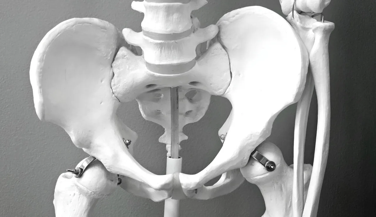
There is more than one route to a pelvic ultrasound, and the sonographer chooses based on the clinical question and your comfort. According to RadiologyInfo, the main approaches are:
- Transabdominal: the probe glides over the lower belly with gel. This wider view usually needs a full bladder, which pushes bowel gas aside and acts as a window to the organs behind it.
- Transvaginal: a slim, covered probe is gently placed inside the vagina for a closer, more detailed view of the uterus and ovaries. Our guide to the transvaginal ultrasound covers this approach in full.
- Transrectal: used mainly in men to look closely at the prostate, the probe is placed into the rectum.
When the study is extended upward to include the liver, kidneys and other upper organs, it is often described as a combined scan. Our guide to the USG abdomen and pelvis explains that wider examination.
Why a doctor orders one
A pelvic ultrasound is a common first line test because it is safe, quick and informative. Typical reasons a clinician might request one include:
- Investigating pelvic pain, pressure or a swelling that can be felt.
- Looking into abnormal or heavy menstrual bleeding.
- Checking the ovaries and uterus for cysts, fibroids or other growths.
- Helping to assess fertility problems or confirm and monitor an early pregnancy.
- Examining the bladder, or in men the prostate, for enlargement or other changes.
- Guiding a procedure such as taking a sample or draining fluid.
Ultrasound often works alongside other tests. If a finding needs more detail, your doctor may add cross sectional imaging such as an MRI scan, and blood flow can be assessed by adding color Doppler ultrasound during the same appointment.
How to prepare

The most important step is to follow the exact instructions your clinic gives you, because preparation differs by the type of scan planned. As a general guide:
- For a transabdominal pelvic scan you are often asked to drink water beforehand and arrive with a comfortably full bladder.
- For a transvaginal or transrectal scan you are usually asked to empty your bladder first.
- Wear loose, comfortable clothing, since you may only need to uncover the lower abdomen or change into a gown.
- Bring your referral, any previous imaging and a list of your medicines.
Tip: If your clinic has not told you whether to arrive with a full or empty bladder, call and ask before you leave home. Getting this wrong is one of the most common reasons a pelvic scan has to be rescheduled or partly repeated.
What happens during the scan
A pelvic ultrasound usually takes about fifteen to thirty minutes. For a transabdominal scan you lie on your back, the sonographer applies warm gel to your lower abdomen, and moves the probe across the skin, pressing gently and changing angle to view each structure. A full bladder can feel uncomfortable during this part, which is normal.
If a transvaginal or transrectal view is needed, the sonographer explains the step first and covers the slim probe with a protective sheath and gel. It is inserted gently and is usually well tolerated, though it can feel slightly uncomfortable. You can ask questions, pause the exam, or request a chaperone at any point. When the scan is finished the gel is wiped off and you can normally return to your usual activities straight away.
Understanding your results
A radiologist reviews the recorded images and writes a report for the doctor who referred you, so you will often not get results during the appointment itself. The report is descriptive: it might note that the uterus and ovaries look normal, describe a simple ovarian cyst, a fibroid, a thickened lining, fluid in the pelvis, or an enlarged prostate. These are observations, not a final diagnosis.
The same wording can mean very different things for two people, because context from your symptoms, age, cycle and blood tests all shape what a finding means. Many findings, such as small simple cysts, are common and harmless, while others need follow up or comparison with an earlier scan. That interpretation is exactly what your clinician is trained to provide.
Important: This article gives general educational information and is not medical advice. It cannot tell you what your own results mean. Please discuss your report, symptoms and any next steps with your referring doctor or a radiologist.
Benefits, limits and safety
A pelvic ultrasound is safe, painless and uses no ionising radiation, so it is well suited to women of childbearing age and to pregnancy, and it can be repeated freely. It is widely available, comparatively inexpensive and gives real time images with good soft tissue detail.
Its limits come mostly from physics. Sound waves are blocked by gas and bone and weaken as they travel through more tissue, so bowel gas or a larger body habitus can reduce clarity on a transabdominal view, which is one reason an internal scan is sometimes added. Ultrasound is also operator dependent, meaning image quality relies on the skill of the sonographer, which is a strong reason to choose an accredited clinic. When ultrasound cannot fully answer the question, an MRI or CT may follow.
How to find a good clinic
When choosing where to have your scan, put quality ahead of the lowest price. Confirm that the clinic is accredited, that scans are performed by qualified sonographers and reported by a radiologist, and that the written report will reach your doctor promptly. Ask whether your images are stored so future scans can be compared. Our guide to how to find and compare ultrasound clinics walks through what to check before you book.
Prices vary by country, provider and whether you pay privately or use insurance, so confirm the fee, exactly what it includes and your coverage before booking. A scan bundled with a radiologist report generally offers better value than a cheaper scan with no interpretation.
Frequently asked questions
Do I need a full bladder for a USG pelvis?
It depends on the type of scan. A transabdominal pelvic ultrasound usually needs a comfortably full bladder to give a clear view, so you are asked to drink water beforehand. A transvaginal or transrectal scan usually needs an empty bladder. Always follow the specific instructions your clinic gives you.
Is a transvaginal ultrasound painful?
Most people find it uncomfortable rather than painful. The probe is slim, covered and lubricated, and the sonographer inserts it gently after explaining the step. You can pause the exam, ask questions or request a chaperone at any time. Tell the sonographer if you feel pain so they can adjust.
Is a pelvic ultrasound safe during pregnancy?
Yes. Ultrasound uses sound waves rather than ionising radiation, so it has no known harmful effects and is used routinely to monitor pregnancy. As with any test, it should still be performed by trained staff for a genuine clinical reason.
How long does the scan take and when will I get results?
The scan itself usually takes about fifteen to thirty minutes. A radiologist then reviews the images and writes a report, so your written result often reaches you a few days later through the doctor who referred you.
What can a pelvic ultrasound detect?
It can show many things, including ovarian cysts, uterine fibroids, changes in the womb lining, an early pregnancy, fluid in the pelvis, and in men an enlarged prostate. What any finding means for you depends on your history and other tests, which is why a doctor must interpret it.
Sources and further reading:
- Pelvic Ultrasound, RadiologyInfo.org for the transabdominal, transvaginal and transrectal approaches, uses and safety.
- Transvaginal Ultrasound, MedlinePlus (US National Library of Medicine) for how the internal scan is performed and what it can show.
- Ultrasound scan, NHS for ultrasound types and why the test uses no radiation.
More guides on Ultrasound Types & Procedures
Have a question or a topic to suggest?
We read every message. Tell us what you would like to see next.
You may also like
CT Imaging Near Me: Compare Clinics
Holding a CT referral and choosing between centers? Use a simple scorecard to compare accreditation, reporting, price, and turnaround instead of picking the first result.
DEXA Scan Near Me: Choose a Clinic
A DEXA scan is one test where the clinic you pick has a lasting effect, because results are compared over years. Learn how to choose a good bone density center.

Ultrasound Locations Near Me
Hospitals, imaging centers, and keepsake studios all advertise ultrasound. Learn to tell a proper diagnostic location from a novelty one before you book.