USG Abdomen & Pelvis: Uses & Results
Why it is ordered, how it works, how to prepare, and what the report means, in plain language.

USG abdomen and pelvis is one of the most commonly ordered scans in medicine, and it often arrives on a request form with almost no explanation. USG simply means ultrasonography: a painless scan that uses high frequency sound waves, not radiation, to look at the organs in your belly and pelvis. This guide explains why the scan is ordered, how it works, how to prepare, and what the report language tends to mean in general terms.
The short version
- A USG of the abdomen and pelvis uses sound waves to image organs such as the liver, gallbladder, kidneys, bladder, uterus, ovaries or prostate.
- It uses no ionizing radiation, which is why it is often the first-line test and is considered safe to repeat.
- Preparation usually means fasting for the abdominal part and a full bladder for the pelvic part, but always follow the exact instructions your clinic gives you.
- A radiologist interprets the images and sends a report to the doctor who ordered the scan. Only that clinician can tell you what your results mean for you.
What USG abdomen and pelvis actually means
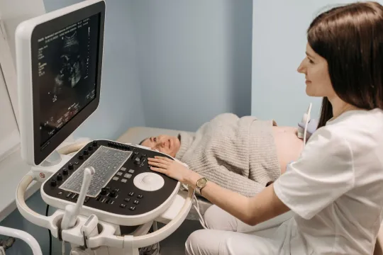
USG is short for ultrasonography, and it is the same technology people mean when they say ultrasound, sonography or a scan. A small handheld device called a transducer is moved across your skin. It sends pulses of high frequency sound into the body and listens for the echoes that bounce back from tissues of different densities. A computer turns those echoes into a live grayscale image on a screen. Because it works with sound rather than X-rays, an ultrasound exam involves no ionizing radiation at all, as the patient resource from RadiologyInfo.org confirms.
An abdomen and pelvis study combines two neighbouring regions into one appointment. The abdominal portion looks at the upper belly organs, while the pelvic portion looks lower down at the bladder and the reproductive organs. Ordering them together is common when symptoms could come from either area, such as unexplained pain, or when a doctor wants a broad first look before deciding on any further imaging.
Why a doctor might order this scan

Doctors reach for an abdominal and pelvic ultrasound in a wide range of situations because it is quick, widely available and free of radiation. Common reasons include the following, though your own reason may differ:
- Investigating abdominal or pelvic pain, bloating or a lump that can be felt.
- Checking the liver, gallbladder and bile ducts for gallstones, fatty change or blockage.
- Looking at the kidneys and bladder for stones, swelling or urine that is not draining.
- Assessing the uterus and ovaries in women, for example when investigating heavy periods, cysts or fertility questions.
- Examining the bladder and prostate in men.
- Following up an abnormal blood test or a finding seen on another scan.
Ultrasound is only one tool in the imaging toolbox. If sound waves cannot reach an area well, or if more detail is needed, your clinician may move on to a CT scan or an MRI scan. Each method has strengths, and the right choice depends on the question the doctor is trying to answer.
What each part of the scan examines
Because the study covers two regions, it helps to know what the sonographer is looking at in each. The exact organs included depend on why your doctor ordered the scan, but a combined abdomen and pelvis study commonly covers the following.
In the abdominal portion, the sonographer typically assesses:
- Liver: size, texture and any masses or signs of fatty change.
- Gallbladder and bile ducts: gallstones, wall thickening or blockage.
- Kidneys: stones, swelling, cysts or obstruction to urine flow.
- Pancreas and spleen: size and obvious abnormalities, when they can be seen clearly.
- Major blood vessels: the aorta and its branches, for example to check for widening.
In the pelvic portion, the focus shifts lower:
- Bladder: filling, wall and how completely it empties.
- Uterus and ovaries in women: fibroids, cysts, the lining and other gynaecological questions.
- Prostate in men: size and outline, often alongside the bladder.
Not every organ is relevant to every patient, and the sonographer follows the specific protocol matched to your referral.
How the scan works, step by step

Knowing the sequence ahead of time takes away most of the anxiety. A typical appointment runs like this:
- You change into a gown or expose the area to be scanned, then lie on an examination couch.
- The sonographer applies a warm water based gel to your skin. The gel removes air gaps so the sound waves can pass into the body cleanly.
- The transducer is pressed gently and moved over the abdomen and pelvis. You may be asked to breathe in and hold, or to roll onto your side, so the organs shift into a better view.
- For the pelvic part, a transabdominal view is taken over a full bladder first. In some cases a transvaginal or transrectal approach gives a clearer internal view.
- The images are captured and the gel is wiped off. Most studies take around 20 to 45 minutes.
Good to know: The gel can feel cool and the transducer may press on a tender area, but the scan should not be painful. Tell the sonographer if anything hurts, because they can adjust their technique.
How to prepare
Preparation is where the abdominal and pelvic parts pull in opposite directions, which is why clear instructions matter. For the abdominal organs, especially the gallbladder, you are usually asked to fast for several hours so the gallbladder is full and the bowel gas is reduced. For the pelvic organs, you are often asked to drink water and arrive with a full bladder, because a full bladder pushes bowel out of the way and acts as a window for the sound waves. RadiologyInfo notes that a pelvic study may require you to drink water beforehand to fill the bladder.
Because these two requirements can seem to conflict, the clinic will give you a tailored plan, often fast from food but keep drinking water. The single most important step is to read and follow the exact letter or message your clinic sends. If anything is unclear, phone them before the day rather than guessing.
Is it safe, and what are the limits
Diagnostic ultrasound has an excellent safety record and has been used for decades, including throughout pregnancy. It uses no ionizing radiation, so there is no radiation dose to weigh up, unlike X-ray or CT, a point the NHS also highlights when explaining why ultrasound is favoured for many scans. The main limitations are practical rather than dangerous: sound waves struggle to pass through gas and bone, so bowel gas or body habitus can obscure some structures, and very deep organs can be harder to see clearly. When that happens, the radiologist will simply recommend a different or additional test.
Understanding the report in general terms
After the scan, a radiologist reviews the images and writes a report for the doctor who referred you. You may see phrases such as unremarkable, meaning nothing abnormal was seen, or descriptions of a cyst, stone, mass or an enlarged organ. It is natural to want to decode these words yourself, but a report is written for another clinician and reads out of context on its own.
A finding is not a diagnosis. The same word can be trivial in one person and important in another, depending on symptoms, history and other test results. That is why the person who ordered the scan is the right one to interpret it for you and to explain any next steps. If you are exploring where to have the scan done, our guides to a clinic with ultrasound near you and to reading a plain abdominal ultrasound can help you know what to ask.
Please note: This article is general educational information, not medical advice. It cannot tell you what your symptoms or your results mean. Always discuss your scan, your report and any next steps with a qualified doctor or your referring clinician.
Frequently asked questions
What is the difference between USG and ultrasound?
There is no difference. USG stands for ultrasonography, which is the same thing as an ultrasound or sonography scan. All three words describe an imaging method that uses high frequency sound waves to create pictures of the inside of the body.
Do I need a full bladder or an empty stomach for a USG of the abdomen and pelvis?
Often both apply to different parts of the exam: an empty stomach for the abdominal organs, and a full bladder for the pelvic organs. Because the exact plan varies, follow the specific fasting and water drinking instructions your clinic gives you, and call them if it is unclear.
Is an abdominal and pelvic ultrasound safe?
Yes. Diagnostic ultrasound uses sound waves and no ionizing radiation, so it carries no radiation dose and is considered very safe, including during pregnancy. It is a painless test, although the transducer may press on a tender area.
How long does the scan take and when will I get results?
The scan itself usually takes about 20 to 45 minutes. A radiologist then reviews the images and sends a report to your referring doctor, which can take anywhere from the same day to several days depending on the clinic. Your doctor will share and explain the results with you.
Can this scan diagnose cancer?
Ultrasound can detect abnormalities such as masses or cysts and can raise or lower suspicion, but on its own it does not confirm or rule out cancer. A diagnosis usually needs further tests, sometimes a biopsy, and is always made by your medical team, not by the scan report alone.
More guides on Ultrasound Types & Procedures
Have a question or a topic to suggest?
We read every message. Tell us what you would like to see next.
You may also like
Abdominal Ultrasound: Uses and Results
A clear guide to the abdominal ultrasound: which organs it checks, why doctors order it, how to prepare and how to understand the report your clinician receives.

Transvaginal Ultrasound: A Guide
A transvaginal ultrasound uses sound waves to produce close-up images of the uterus and ovaries. Here is why it is ordered, exactly what happens during the scan, how to prepare, and what the results can show.
Ultrasound: Uses, Procedure & Results
An ultrasound uses sound waves, not radiation, to image soft tissue in real time. This guide covers why scans are ordered, how they work, how to prepare, and how results are interpreted, with a reminder to discuss findings with your doctor.