Transvaginal Ultrasound: A Guide
Why an endovaginal scan is ordered, what actually happens step by step, how to prepare, and how to feel comfortable and in control.

A transvaginal ultrasound, also called an endovaginal scan, produces close-up images of the uterus, ovaries, and other pelvic structures using sound waves rather than radiation. It is a common and safe test, and understandably a lot of people feel nervous before their first one. This guide explains why it is ordered, exactly what happens during the procedure, how to prepare, and why only your doctor can tell you what the results mean.
The short version
- A slim transducer is gently placed in the vagina to image the uterus, endometrium, and ovaries up close.
- It uses sound waves, not radiation, and is considered very safe.
- You usually empty your bladder first, the opposite of a scan done over the abdomen.
- A radiologist interprets the images and reports to your doctor, who explains what they mean for you.
What a transvaginal ultrasound is
Transvaginal ultrasound is a type of pelvic ultrasound. Instead of scanning through the abdominal wall, a thin transducer is placed a short way into the vagina, which brings it much closer to the uterus and ovaries. That proximity gives clearer, more detailed images than an abdominal scan can, especially of the endometrium (the lining of the uterus) and the ovaries.
Like all ultrasound, it works by sending high-frequency sound waves into the body and building a picture from the echoes that bounce back. As the NHS notes, ultrasound uses no radiation, unlike x-rays or CT scans. For a general grounding in how these scans work, see our overview of ultrasound imaging.
Why a doctor orders one

A transvaginal ultrasound is ordered when a doctor needs a detailed look at the female reproductive and urinary structures. According to RadiologyInfo.org, common reasons include:
- Investigating pelvic pain or abnormal vaginal bleeding.
- Looking into fertility problems or monitoring treatment.
- Assessing ovarian cysts, uterine fibroids, or polyps.
- Checking the endometrium, for example after bleeding beyond menopause.
- Confirming and dating a very early pregnancy, when it is too small to see well on an abdominal scan.
Because the transducer sits so close to the organs, it is often the preferred way to examine early pregnancy and subtle changes in the uterine lining. It is also frequently used to locate an intrauterine device, to guide fertility treatment by tracking follicles, and to help investigate the cause of recurrent miscarriage. In each case the scan gives information that guides the next step, rather than a diagnosis on its own.
How the procedure works, step by step
Knowing exactly what happens tends to ease the nerves. The scan is usually quick and should not be painful.
- You empty your bladder, then undress from the waist down and lie back with your knees bent, often with your feet in supports.
- The sonographer covers a slim transducer with a protective sheath and lubricating gel.
- The transducer is gently inserted a few centimetres into the vagina. RadiologyInfo.org notes its tip is smaller than the standard speculum used for a Pap test.
- The sonographer angles it slightly to view the uterus and each ovary, capturing images as they go. Colour Doppler may be used to show blood flow.
- The transducer is removed, and the whole appointment typically takes around 30 minutes.
You may feel mild pressure but it should not be significantly uncomfortable. If it hurts, tell the sonographer, who can adjust what they are doing.
How to prepare: For a transvaginal scan you usually empty your bladder just before, which is the opposite of an abdominal pelvic scan where a full bladder helps. Wear something easy to change out of. If you are having your period, the scan can often still go ahead, but call the clinic if you are unsure, and mention any tampon so it can be removed first. You can ask for a chaperone at any time.
Is it safe?
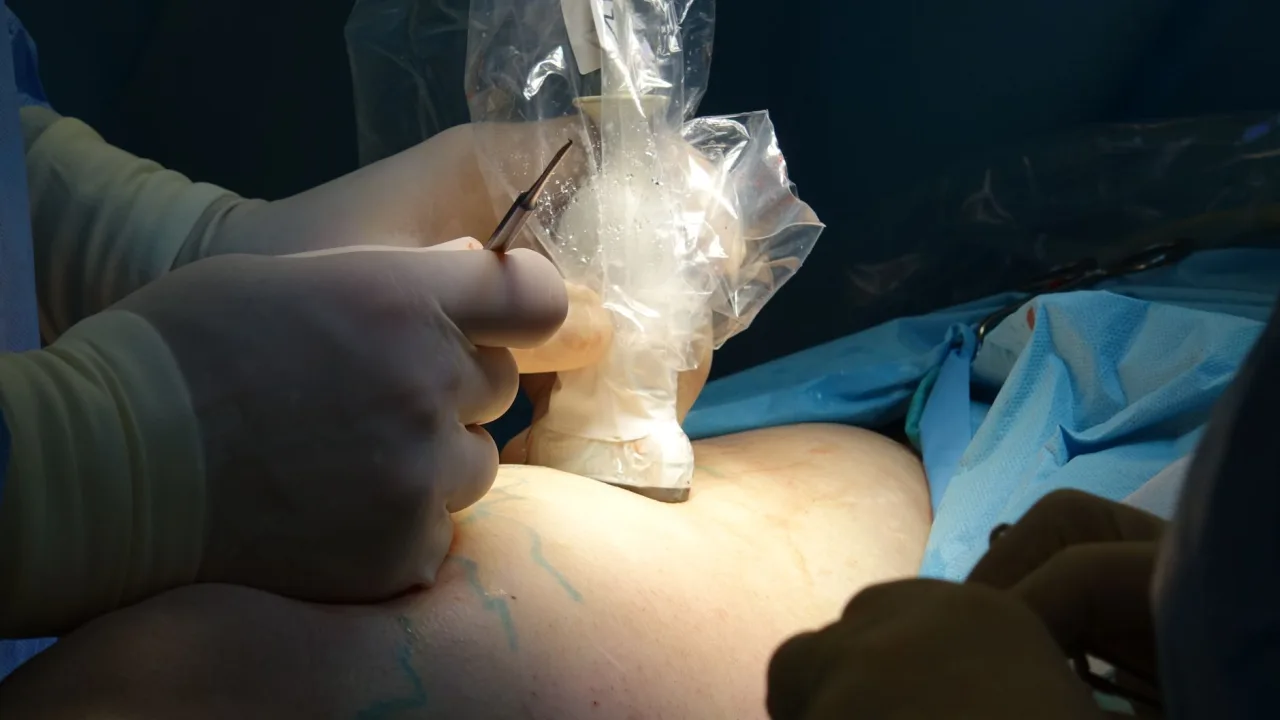
Yes. Diagnostic ultrasound has no known harmful effects on the body and uses no ionising radiation, which is why it is used routinely in pregnancy. The scan is minimally invasive: the transducer sits in the vagina but does not enter the uterus. Mild pressure is the most anyone usually feels. Because a new sheath and fresh gel are used each time, infection risk is very low.
Because the transducer is cleaned and disinfected between patients and covered with a single-use sheath, cross-infection is very unlikely in an accredited clinic. If you have a latex allergy, tell the clinic beforehand so a suitable sheath can be used. And if inserting the transducer is too uncomfortable, an abdominal scan may be an alternative, though it gives less detail of the uterus and ovaries. Our guide to abdominal ultrasound explains that approach.
Feeling comfortable and in control
It is completely normal to feel awkward or anxious about a transvaginal scan, and a good clinic expects that. You are in control throughout: you can ask questions, ask for a chaperone regardless of the sonographer's gender, and ask the sonographer to pause or stop at any point. Many people find the scan is far less uncomfortable than they feared once it is under way.
A few small things can help. Wear clothing you can change out of easily, arrive with an empty bladder as advised, and let the clinic know in advance if you have a history that makes internal examinations difficult, such as vaginismus or previous trauma, so they can plan a gentler approach or discuss alternatives. You never have to proceed if you do not want to, and choosing an abdominal scan instead is always a reasonable option to raise.
What the results mean
A radiologist reviews the images and sends a report to the doctor who ordered the scan. That doctor then explains the findings in the context of your symptoms, history, and any other tests. The scan can show the size and shape of the uterus, the thickness of the endometrium, the ovaries and any cysts, and, in pregnancy, an early gestational sac and heartbeat.
Timing within your menstrual cycle can affect what the scan shows, because the endometrium naturally thickens and thins through the month and ovarian follicles change in size. For this reason a doctor sometimes asks for the scan on a particular day of your cycle, and the same measurement can be normal at one point and worth a closer look at another. This is one more reason the raw numbers on a report are not meant to be read in isolation.
This is general information, not medical advice. Findings like a cyst, a fibroid, or a particular lining thickness can be completely normal or need follow-up, depending entirely on your situation. Do not try to interpret your own images or report. Always discuss the results with the doctor who ordered the scan or a specialist they refer you to.
Related scans and next steps
Transvaginal ultrasound often works alongside other tests. In early pregnancy it may be followed by later scans, and our guide to a pregnancy sonogram week by week sets out what to expect as pregnancy progresses. If you are arranging the scan yourself, our guide to a private ultrasound near you explains how to choose a good clinic. And if the people and skills behind these scans interest you, our sonography career guide looks at how sonographers train.
Frequently asked questions
Does a transvaginal ultrasound hurt?
It should not be painful. Most people feel mild pressure as the slim transducer is inserted a few centimetres. RadiologyInfo.org notes the tip is smaller than the speculum used for a Pap test. If you feel pain, tell the sonographer, who can adjust the angle or stop.
Do I need a full bladder for a transvaginal ultrasound?
No. For a transvaginal scan you usually empty your bladder just before the exam. This is the opposite of an abdominal pelvic ultrasound, where a full bladder helps push the bowel aside and improve the view. The clinic will confirm what to do when you book.
Can I have the scan during my period?
Often yes, a transvaginal ultrasound can still be done during a period, though you may prefer to reschedule for comfort. Any tampon will need to be removed first. If you are unsure, call the clinic, as timing sometimes matters for what the scan is checking.
Is a transvaginal ultrasound safe in pregnancy?
Yes. Ultrasound uses sound waves, not radiation, and has no known harmful effects, which is why it is used routinely in pregnancy. In early pregnancy a transvaginal scan often gives a clearer view than an abdominal one because the transducer sits closer to the uterus.
Who tells me what the results mean?
A radiologist interprets the images and reports to the doctor who ordered the scan, who then explains the findings to you. Results such as a cyst or a certain lining thickness can be normal or need follow-up depending on your situation, so always discuss them with your doctor.
More guides on Ultrasound Types & Procedures
Have a question or a topic to suggest?
We read every message. Tell us what you would like to see next.
You may also like
Ultrasound: Uses, Procedure & Results
An ultrasound uses sound waves, not radiation, to image soft tissue in real time. This guide covers why scans are ordered, how they work, how to prepare, and how results are interpreted, with a reminder to discuss findings with your doctor.
Abdominal Ultrasound: Uses and Results
A clear guide to the abdominal ultrasound: which organs it checks, why doctors order it, how to prepare and how to understand the report your clinician receives.

Pregnancy Sonogram: Week by Week
A reassuring, week by week look at pregnancy ultrasound scans, including what the dating and anomaly scans check, how to prepare and how results are shared.