Chest X-Ray: Uses, Procedure & Results
What a chest X-ray shows, the small radiation dose involved, how to prepare, and how a radiologist reads the image.
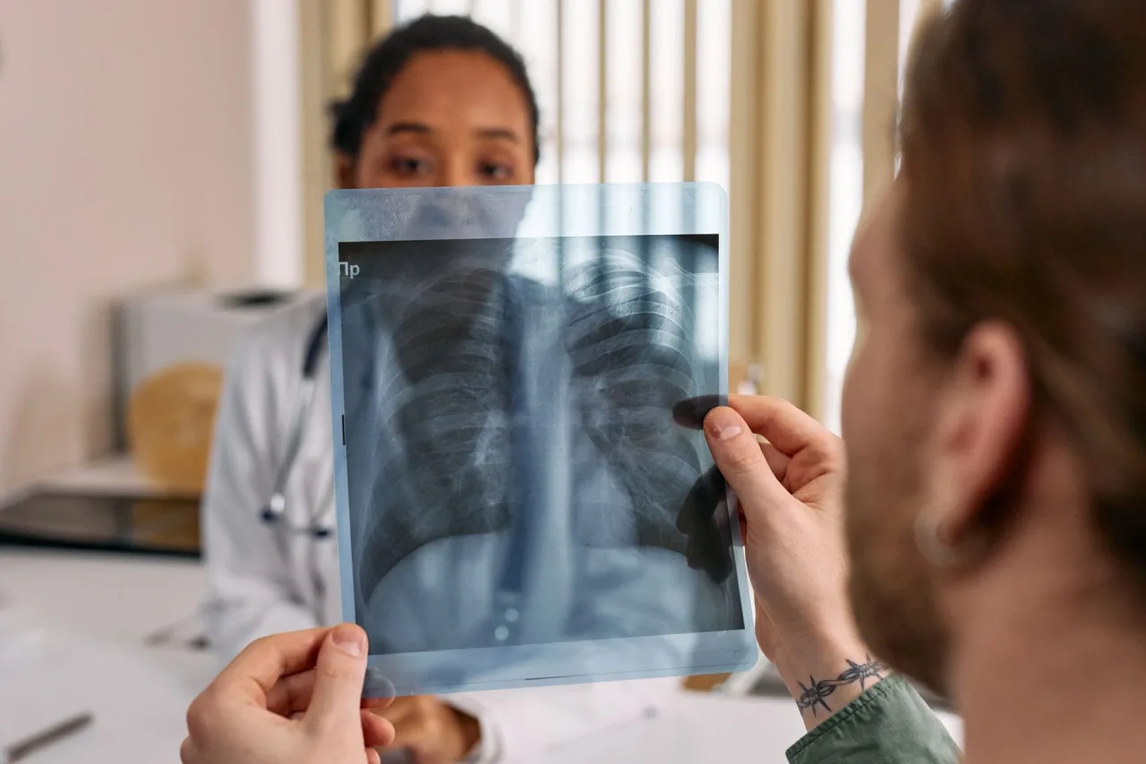
Chest radiography, better known as a chest X-ray, is the most common imaging test in medicine. This guide explains what a chest X-ray is, why it is ordered, how it works, how to prepare, the small radiation dose involved, and how a radiologist reads the result.
If your doctor has asked for a chest X-ray, it is usually a quick, painless step that takes only a few minutes. Still, it helps to understand what the scan can show, what the radiation exposure really amounts to, and why the person taking the image cannot simply tell you the answer on the spot. This article covers all of that in clear, honest terms.
The short version
- A chest X-ray uses a very small dose of ionizing radiation to image the heart, lungs, airways, and chest bones.
- It is the most commonly performed diagnostic X-ray and needs no special preparation.
- The radiation dose is low, but always tell staff if you are or might be pregnant.
- A radiologist interprets the image and reports to your doctor, who explains what it means for you.
What chest radiography is
A chest X-ray produces images of the inside of the chest, including the heart, lungs, airways, blood vessels, and the bones of the spine and chest. According to RadiologyInfo.org, the chest X-ray is the most commonly performed diagnostic X-ray examination. It uses a very small dose of ionizing radiation to create the picture.
The physics is simple to picture. X-rays pass through the body and are absorbed in different amounts by different tissues. Dense structures such as bone absorb more and appear white, while air-filled lungs let more radiation through and appear dark. The result is a single greyscale snapshot that a trained eye can read in remarkable detail. Chest radiography is one part of the broader field of radiology, and it is often the first imaging test before more detailed scans.
Why your doctor might order one

Doctors use chest X-rays to evaluate symptoms and monitor known conditions. Common reasons include the following.
- Breathing problems: shortness of breath, a persistent cough, or chest pain.
- Suspected infection: pneumonia or other lung infections, especially with fever.
- Heart and lungs: signs of heart failure, fluid around the lungs, or an enlarged heart.
- Chronic disease: monitoring conditions such as emphysema, or checking for lung cancer.
- Checking devices: confirming the position of a tube, line, or pacemaker.
A chest X-ray is often a starting point. If it raises a question that needs more detail, your doctor may order a CT scan of the chest, which gives a far more detailed, cross-sectional view. For a deeper look at how chest imaging findings are described and followed up, see our companion guide on chest radiology.
How to prepare
Preparation could hardly be simpler. A chest X-ray requires no special preparation, no fasting, and no injections in most cases. You will be asked to remove jewellery, glasses, and any metal objects around the chest and neck, and often to change into a gown, because metal and dense fabric can show up on the image and obscure the view.
- Remove metal: necklaces, some clothing with metal fasteners, and items in chest pockets.
- Tell staff about pregnancy: women should always tell their doctor and the radiographer if they are or might be pregnant.
- Follow positioning cues: you may be asked to take a deep breath and hold it, which inflates the lungs for a clearer image.
What happens during the scan
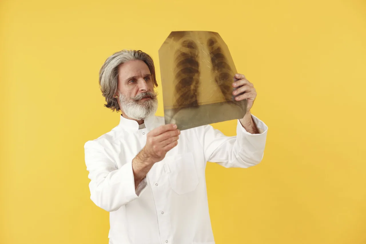
The examination is fast and painless. You usually stand with your chest against a flat image detector, though it can be done sitting or lying down if you are unwell. The radiographer positions you, then steps behind a screen or leaves the room to take the exposure. RadiologyInfo notes this is standard practice, because the staff take many images each day and keep their own cumulative exposure as low as possible, while your single exposure is very small.
Most chest X-rays include two views, one from the back and one from the side, taken while you hold a deep breath for a second or two. Holding your breath inflates the lungs and keeps the chest still, which sharpens the image and helps the radiologist see fine detail. The actual exposure lasts a fraction of a second. The whole appointment, including positioning and changing, is typically over within about ten minutes, and there is no recovery time afterward: you can eat, drive, and return to normal activity straight away.
Radiation dose and safety
The word radiation understandably makes people cautious, so it helps to keep the amount in perspective. A chest X-ray uses a very small dose of ionizing radiation. The NHS describes an X-ray as a test that uses a small amount of radiation and notes that X-rays are usually very safe, with serious complications being rare. Importantly, no radiation stays in your body after the exam is over.
For any medically indicated chest X-ray, the benefit of the information it provides far outweighs the tiny risk from the dose. The main precaution concerns pregnancy: if you are or might be pregnant, tell your care team so they can decide whether to proceed, shield you, or choose a test without radiation such as ultrasound. Doctors follow the principle of using the lowest dose that still answers the clinical question.
Important: Always disclose a known or possible pregnancy before any X-ray. Your care team will weigh the benefits and risks and may use shielding or an alternative test.
Understanding your results
Here is the honest reality of results: producing the image and interpreting it are two different skills. A radiologist reviews your chest X-ray and writes a report, which goes to the doctor who referred you. That doctor then explains what it means in the context of your symptoms, history, and any other tests.
Report language such as "opacity," "infiltrate," "effusion," or "unremarkable" can be hard to interpret alone, and some findings are old, stable, or harmless. A single image is a snapshot, and its meaning often depends on comparison with previous films or further tests. This is exactly why you should bring questions to your clinician rather than trying to decode the wording yourself.
Note: This article is general information, not medical advice, and it cannot tell you what your own X-ray shows. Please discuss your report and any symptoms with your referring doctor or a qualified radiologist.
Who takes and reads the image
Two professionals are usually involved. A radiographer, sometimes called an X-ray technologist, positions you and captures the image using the equipment safely. A radiologist, a doctor specialised in interpreting medical images, reads the film and writes the diagnostic report. If a career in taking these images interests you, our guide to becoming an X-ray technologist outlines the training and daily work involved.
Typical costs
Chest X-rays are among the least expensive imaging tests, but the price still varies by country, provider, and insurance. In many public health systems a medically necessary chest X-ray is free at the point of care. Where you pay privately, the cost is often modest compared with CT or MRI, though the exact figure depends on the facility and whether a specialist reading fee is added. Treat any figure you see as a rough range only. Confirm the price with the clinic and check coverage with your insurer before booking.
Frequently asked questions
Is a chest X-ray safe?
Yes, for medically indicated exams the risk is very small. A chest X-ray uses a very small dose of ionizing radiation, and no radiation stays in your body afterward. The information it provides for a genuine medical reason far outweighs the tiny risk from the dose.
Do I need to prepare for a chest X-ray?
No special preparation is needed. You do not need to fast. You will be asked to remove jewellery and metal objects near the chest, and often to change into a gown, because metal can obscure the image. You may be asked to take a deep breath and hold it during the exposure.
Can I have a chest X-ray if I am pregnant?
Always tell your doctor and the radiographer if you are or might be pregnant. They will weigh the benefits and risks, and may use shielding, delay the test, or choose an alternative such as ultrasound that does not use radiation.
How long does it take and when will I get results?
The appointment itself usually takes under ten minutes. A radiologist then interprets the image and reports to your referring doctor, who gives you the results. Timing ranges from the same day for urgent cases to several days for routine ones.
What is the difference between a chest X-ray and a chest CT?
A chest X-ray is a single, quick, low-dose image and is often the first test. A CT scan uses more radiation to build detailed cross-sectional images and is used when a doctor needs a closer look at something the X-ray raised. Your doctor decides which is appropriate for your situation.
More guides on Ultrasound & Radiology (General)
Have a question or a topic to suggest?
We read every message. Tell us what you would like to see next.
You may also like

Chest X-Ray: A Patient's Guide
What chest radiology and the chest X-ray involve: what the scan shows, how it is done, radiation safety, and what the report actually means.

X-Ray Tech: Uses, Procedure & Results
A clear guide to X-rays: why they are ordered, how the test works, radiation safety, who performs and reads them, and how to think about your results.

CT Scan: Uses, Procedure and Results
A CT scan, or CAT scan, uses x-rays and a computer to create detailed cross-sectional images of the body. Here is why doctors order one, how the procedure works, what contrast dye does, and how the radiation compares.