Ultrasound Abdomen and Pelvis
An ultrasound of the abdomen and pelvis is a single appointment that looks at two neighbouring regions of the body using sound waves.
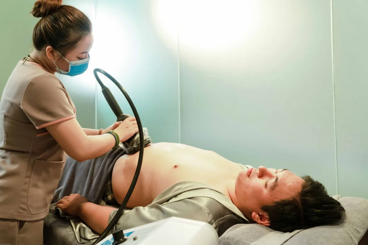
An ultrasound of the abdomen and pelvis is a single appointment that looks at two neighbouring regions of the body using sound waves. It is a common, radiation-free way to investigate pain, check organs, and follow up abnormal blood tests. This guide explains what the scan covers, why the two areas are often examined together, and how to prepare for the slightly contradictory bladder instructions that can catch people out.
The short version
- A combined abdomen and pelvis ultrasound checks upper abdominal organs and the pelvic organs in one visit.
- It uses sound waves, not radiation, so it is safe and painless for most people.
- Preparation can seem to conflict: you may be asked to fast for the abdominal part yet arrive with a full bladder for the pelvic part. Always follow your clinic's exact instructions.
- A sonographer captures the images, but a radiologist interprets them, and only your doctor can explain what the results mean for you.
What the scan covers
The abdomen and the pelvis sit next to each other, and a combined scan gives a broad view across both. The RadiologyInfo abdominal ultrasound page explains that an abdominal ultrasound evaluates organs such as the kidneys, liver, gallbladder, bile ducts, pancreas, spleen, and the abdominal aorta. The RadiologyInfo pelvic ultrasound page adds that a pelvic scan examines the bladder and, depending on your anatomy, the uterus, ovaries, and fallopian tubes, or the prostate and seminal vesicles.
Combining the two is useful when symptoms could arise from either region, such as generalised abdominal pain, or when a clinician wants a fuller survey in one sitting. If your referral mentions only one area, our focused explainers on abdominal ultrasound and the broader field of diagnostic ultrasound go into more depth.
Why it is ordered

A doctor may request a combined abdomen and pelvis ultrasound to investigate a range of concerns, including:
- Abdominal or pelvic pain that has not been explained.
- Suspected gallstones, kidney stones, or urinary problems.
- Abnormal liver or kidney blood tests.
- A lump, swelling, or a mass found on examination or another scan.
- Follow-up of a known cyst, fibroid, or other finding.
Because ultrasound shows soft tissue and blood flow well and uses no radiation, it is frequently the first imaging test chosen for these questions. If the results raise further questions, a doctor may then consider other tests, such as a CT scan of the abdomen and pelvis or an MRI scan.
How to prepare
Preparation is the part people most often get wrong, because the two regions can call for opposite things. Your clinic will give exact instructions, but common patterns are:
- Fasting for the abdominal part: RadiologyInfo notes that your doctor will tell you whether to refrain from eating or drinking beforehand. Fasting for several hours reduces gas and empties the gallbladder so it can be seen clearly.
- A full bladder for the pelvic part: RadiologyInfo notes you may be asked to drink water to fill your bladder, which improves the view of the pelvic organs during a scan through the abdomen.
- Practical steps: wear loose, comfortable clothing and leave valuable jewellery at home.
Tip: If your instructions say both fast and arrive with a full bladder, that is normal for a combined scan. It usually means no food for several hours but drinking water shortly before the appointment. If anything is unclear, phone the clinic and confirm rather than guessing.
How the scan is performed

The appointment is straightforward and usually painless. A typical sequence is:
- You lie on your back on an examination couch, and the sonographer exposes the abdomen and lower abdomen.
- A small amount of warm gel is applied to the skin so the probe makes good contact.
- The sonographer moves a handheld probe across the abdomen and pelvis, sometimes asking you to breathe in, hold your breath, or turn slightly.
- For some pelvic questions, a transvaginal or transrectal approach gives a closer view, and this would be explained and offered separately.
The whole examination often takes 20 to 45 minutes depending on how much needs to be covered. If a closer internal view of the pelvis is suggested, our guide to the transvaginal ultrasound explains that step in detail.
Is it safe?
Ultrasound is considered very safe. RadiologyInfo states that ultrasound is safe, noninvasive, and does not use ionising radiation, and that standard diagnostic ultrasound has no known harmful effects on humans. That safety profile is a major reason it is used so widely, including in pregnancy. It does have limits: ultrasound cannot see well through gas or bone, so bowel gas can obscure parts of the abdomen, and imaging can be harder in larger patients.
What the results mean
After the scan, a radiologist reviews the images and writes a report for the doctor who referred you. You may not get answers on the day, because the sonographer performing the scan is often not the person who issues the final interpretation. The report describes what was seen, such as the size and appearance of organs, any stones, cysts, or masses, and whether anything needs further tests.
It is important not to interpret your own images or a brief comment during the scan. A finding that sounds worrying in isolation may be minor in context, and a normal-looking scan does not rule out every condition. Your doctor pulls the report together with your symptoms, examination, and blood tests to decide what happens next, which might be reassurance, treatment, or another scan. If cost is a factor in where you have the scan, our guide to a diagnostic center near you covers what to ask about.
Please note: This article is general educational information, not medical advice. It cannot tell you what your own scan shows or what any finding means for your health. Please discuss your report and any symptoms with your doctor or the radiology team.
Frequently asked questions
What organs does an abdomen and pelvis ultrasound show?
The abdominal part typically looks at the kidneys, liver, gallbladder, bile ducts, pancreas, spleen, and the abdominal aorta. The pelvic part looks at the bladder and, depending on your anatomy, the uterus and ovaries or the prostate. The exact organs depend on why the scan was ordered.
Do I need a full bladder and to fast at the same time?
For a combined scan this combination is common. It usually means not eating for several hours to see the abdominal organs clearly, then drinking water shortly before the appointment so the bladder is full for the pelvic views. Follow the exact instructions your clinic gives you, and call them if anything is unclear.
Does the scan use radiation?
No. Ultrasound uses sound waves, not ionising radiation, which is one reason it is often the first imaging test chosen for abdominal and pelvic questions. It is considered safe and has no known harmful effects at standard diagnostic levels.
How long does an abdomen and pelvis ultrasound take?
It often takes around 20 to 45 minutes, depending on how much needs to be examined and whether any closer internal views of the pelvis are needed. The sonographer may ask you to change position or hold your breath at times.
When will I get my results?
Usually not on the day. A radiologist reviews the images and sends a report to the doctor who referred you, who then explains what it means in the context of your symptoms and other tests. If you have not heard back within the expected time, contact the referring clinic.
More guides on Ultrasound & Radiology (General)
Have a question or a topic to suggest?
We read every message. Tell us what you would like to see next.
You may also like

5 Week Ultrasound
A 5 week ultrasound is one of the earliest scans in pregnancy, usually arranged for a specific reason rather than as routine care. At this very early stage the goal is modest

4 Week Ultrasound
A 4 week ultrasound is about as early as a pregnancy scan can be attempted, and at this point it is often too soon to see much at all. Many people search for it hoping for reassurance right after a positive test

Abdomen and Pelvis Scan
An abdomen and pelvis scan is not a single test. The phrase can mean an ultrasound, a CT scan, or an MRI, and the three work in very different ways, need different preparation