CT Scan Abdomen and Pelvis
A CT scan of the abdomen and pelvis uses x-rays and a computer to build detailed cross-sectional pictures of the organs in your belly and lower trunk.
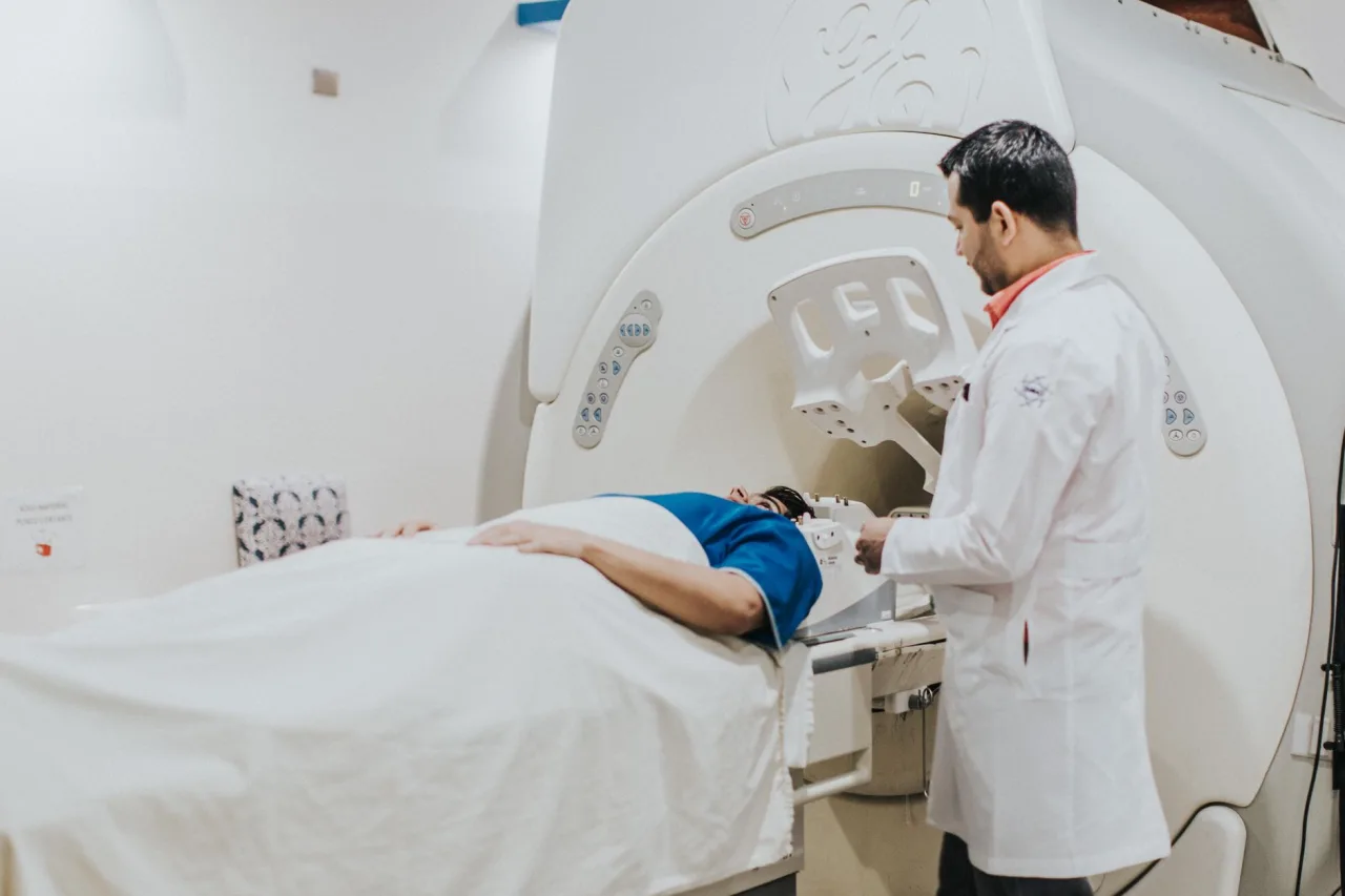
A CT scan of the abdomen and pelvis uses x-rays and a computer to build detailed cross-sectional pictures of the organs in your belly and lower trunk. It is one of the most common ways to investigate abdominal pain and to check organs such as the liver, kidneys, bowel, and bladder. This guide explains why it is ordered, how the procedure works, what contrast dye does, the radiation involved, and why only a doctor can tell you what your results mean.
The short version
- This scan images the abdomen and pelvis together, covering organs from below the chest down to the hips.
- It is quick and painless, and contrast dye is often used to make organs and blood vessels stand out.
- CT uses more radiation than a plain x-ray, so it is ordered when the detail it gives outweighs that small added risk.
- A radiologist interprets the images and reports to your doctor. Never try to read the scan yourself.
What a CT scan of the abdomen and pelvis is
A CT scanner is a doughnut-shaped machine with an x-ray tube that rotates around you, taking many measurements from different angles. A computer combines them into cross-sectional images, often called slices, that can be stacked or rebuilt into three-dimensional views. When the abdomen and pelvis are scanned together, the study covers the liver, gallbladder, spleen, pancreas, kidneys, adrenal glands, bowel, bladder, and the reproductive organs, along with the blood vessels and lymph nodes around them.
According to RadiologyInfo.org, CT of the abdomen and pelvis provides greater clarity than a plain x-ray, especially for soft tissues and blood vessels, and can be reformatted into multiple planes. For a broader look at how CT sits among imaging tests, see our overview of radiology and our general guide to the CT scan.
Why a doctor orders this scan
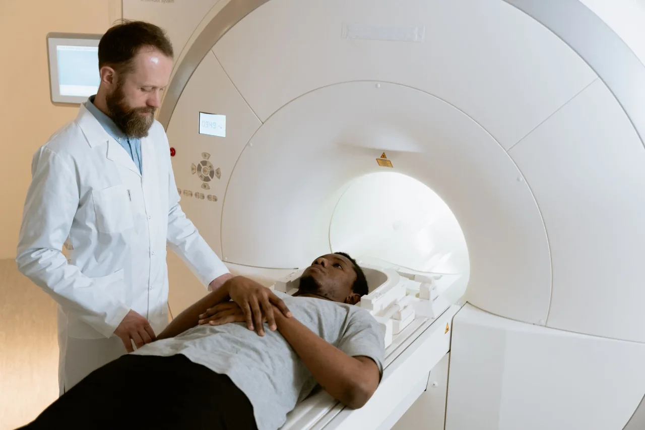
The abdomen and pelvis contain many organs, so this combined scan is a versatile tool. RadiologyInfo.org lists common reasons a doctor requests one, including:
- Finding the cause of abdominal or pelvic pain.
- Investigating infections such as appendicitis, diverticulitis, or an abscess.
- Assessing inflammatory bowel disease and pancreatitis.
- Detecting and staging cancers of the abdominal and pelvic organs.
- Looking for kidney stones and urinary tract problems.
- Assessing injuries after trauma and planning or checking surgery.
Because CT is fast and detailed, it is often the test chosen when someone is acutely unwell and doctors need answers quickly. For comparison, an ultrasound of the abdomen and pelvis uses no radiation and is often the first test for many problems, while CT is used when more detail is needed.
How the procedure works, step by step
The scan itself usually takes only seconds to a few minutes, though the appointment is longer because of preparation.
- You may be asked to fast for a few hours beforehand, and to change into a gown and remove metal items.
- If contrast is used, you may drink a contrast liquid in advance, and a small cannula may be placed in a vein for an injected dye.
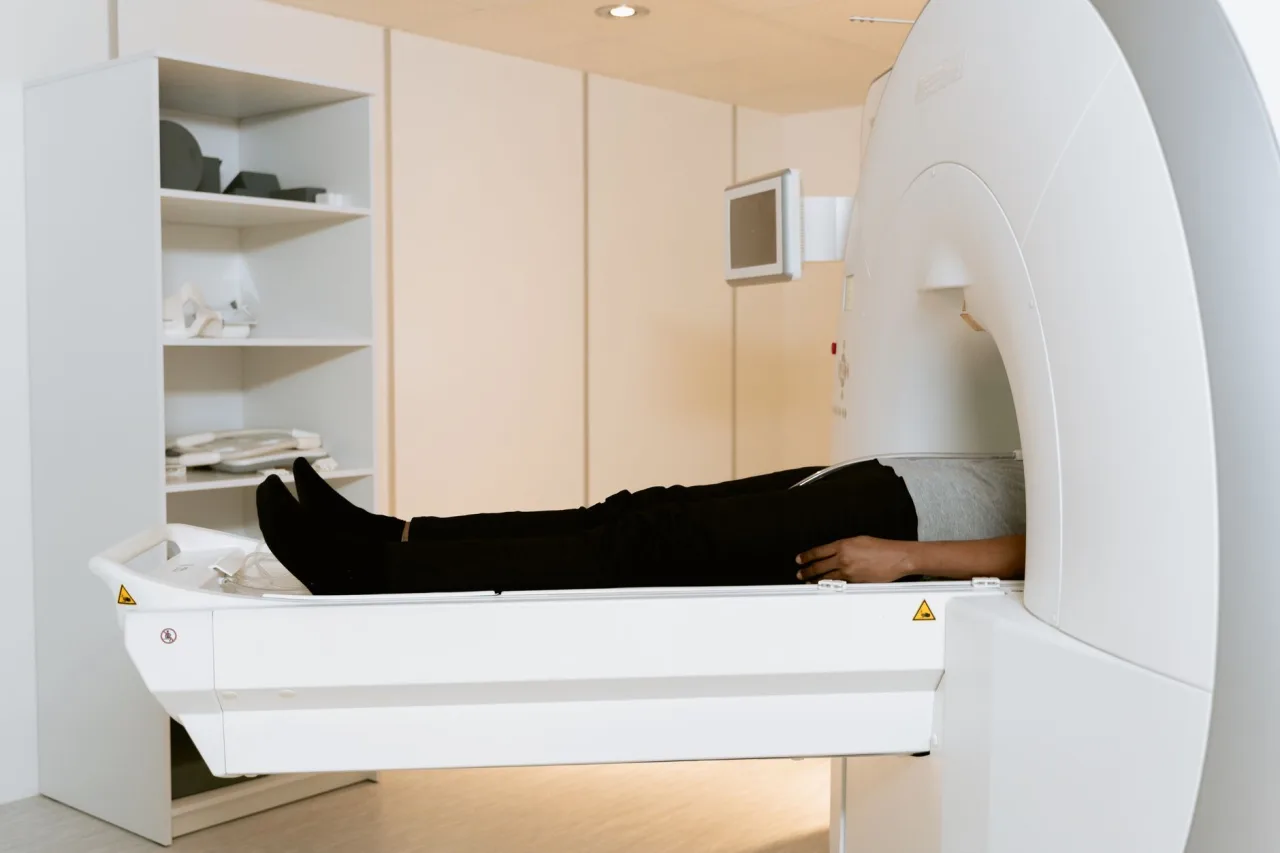
- You lie on your back on a motorised table that slides slowly through the scanner ring.
- You may be asked to hold your breath briefly so the images are sharp, while the technologist watches from an adjoining room.
The scanner is an open ring rather than an enclosed tunnel, so it feels far less confined than an MRI scan. You will not feel the x-rays, though intravenous contrast can cause a brief warm flush or metallic taste that passes quickly.
Contrast dye: what it does
Contrast medium is a dye that makes organs, blood vessels, or the gut stand out more clearly on the images. As RadiologyInfo.org notes, abdominal and pelvic CT often uses an iodine-based contrast, and the risk of a serious allergic reaction to it is extremely rare. Contrast can be swallowed, injected into a vein, or occasionally given into the back passage, and the body clears it naturally afterwards.
Important: Tell the clinic in advance if you have had a reaction to contrast before, have kidney problems, take diabetes medicine, or are pregnant or breastfeeding. Patients with a known allergy may be given preventive medication first. If there is any chance you are pregnant, say so, because CT is generally avoided in pregnancy unless it is essential.
Radiation: keeping it in proportion
A CT of the abdomen and pelvis uses more radiation than a plain x-ray because it takes many images across a large area of the body. RadiologyInfo.org lists a CT of the abdomen and pelvis at roughly 7.7 millisieverts, compared with about 0.1 millisieverts for a single chest x-ray and around 3 millisieverts of natural background radiation a year. No radiation stays in your body after the scan.
Doctors weigh the benefit of the scan against that small risk and use the lowest dose that still answers the question, following the ALARA principle, meaning as low as reasonably achievable. For someone who is unwell, the information a CT provides can be genuinely important, and the small radiation risk is usually far outweighed by an accurate, fast diagnosis. The aim is to make sure each scan is justified and not simply repeated when an earlier scan or an ultrasound would answer the question.
What your results mean
After the scan, a radiologist studies the images and writes a report for the doctor who ordered the test. That doctor then explains the findings in the context of your symptoms and history. A result described as a finding is not automatically serious, and many incidental findings, meaning things noticed by chance, turn out to be harmless.
How quickly you get results depends on the setting. In an emergency, a radiologist may review the images within the hour, while a routine outpatient report can take several days to reach your doctor. It is natural to feel anxious in the gap, but try not to draw conclusions from the images or an online portal before your doctor has explained them. Radiology reports are written for clinicians and often use cautious, technical language that can sound more alarming than the situation warrants.
This is general information, not medical advice. Abdominal and pelvic CT images are complex, and the same appearance can mean very different things depending on your situation. Do not attempt to interpret your own scan or report. Always discuss the findings with the doctor who ordered the scan or a specialist they refer you to.
Cost and where to have the scan
What a CT of the abdomen and pelvis costs varies widely by country, provider, and whether contrast is used, and by whether it is covered by insurance or a public health system. The same scan can differ by a large amount between facilities, so it is always worth confirming both the price and your coverage with the clinic and your insurer before booking. Our detailed guide to the price of a CT scan breaks down realistic ranges and how to ask for a quote.
Choosing an accredited clinic matters more than choosing the closest one. Our guide to finding a CT scan clinic near you explains what to check before you book.
Frequently asked questions
What does a CT scan of the abdomen and pelvis show?
It shows the organs of the belly and lower trunk, including the liver, kidneys, pancreas, spleen, bowel, bladder, and reproductive organs, along with nearby blood vessels and lymph nodes. It is used to investigate pain, infection, cancer, kidney stones, and injuries. A radiologist interprets what the images show.
Do I need to prepare for the scan?
Often yes. You may be asked to fast for a few hours, to drink a contrast liquid beforehand, and to remove metal items. The clinic gives you specific instructions when you book. Tell them about allergies, kidney problems, diabetes medicine, and any chance of pregnancy.
Is the radiation dangerous?
A CT of the abdomen and pelvis uses more radiation than a plain x-ray, roughly 7.7 millisieverts, but it is considered low risk when the scan is medically justified. Having many scans over time carries a very small increase in cancer risk, so doctors order CT only when the benefit outweighs it.
How is it different from an ultrasound of the same area?
An ultrasound uses sound waves and no radiation and is often the first test, while CT uses x-rays to give more detailed cross-sectional images. Doctors choose between them based on the question. CT is frequently used when more detail is needed or in an emergency.
Who tells me the results?
A radiologist interprets the images and sends a report to the doctor who ordered the scan, and that doctor explains what the results mean for you. You should not rely on reading the images or report yourself, because interpretation depends on your full clinical picture.
More guides on MRI, CT & X-Ray
Have a question or a topic to suggest?
We read every message. Tell us what you would like to see next.
You may also like
L Spine X-Ray
An L spine x-ray, short for lumbar spine x-ray, is a quick imaging test that takes pictures of the five bones in your lower back. Doctors use it to look for fractures, alignment problems, arthritis

Foot X-Ray Normal
A foot x-ray is a quick, painless test that shows the many small bones and joints of the foot. If you have searched for foot x-ray normal, you are probably trying to understand what a normal result actually means
MRI Center
An MRI center is a facility, either inside a hospital or as a standalone clinic, that performs magnetic resonance imaging. If your doctor has referred you for an MRI, knowing what happens at one of these centers