Fluoroscopy
Fluoroscopy is a form of medical imaging that turns X-rays into a live, moving picture of the inside of your body.
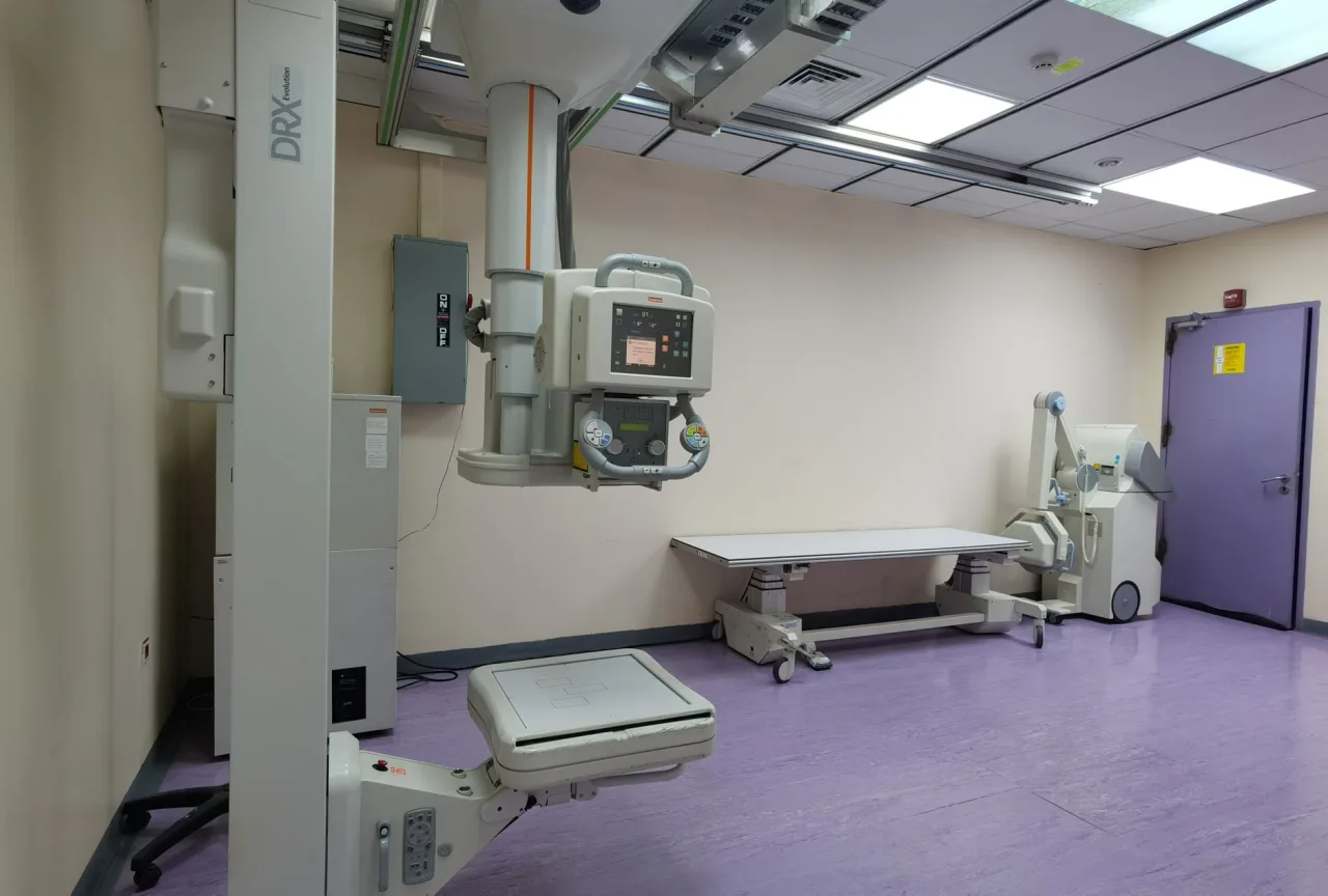
Fluoroscopy is a form of medical imaging that turns X-rays into a live, moving picture of the inside of your body. Instead of a single still photo, your care team watches organs, joints, blood vessels or swallowed contrast move in real time. This guide explains what fluoroscopy is, why it is ordered, how the exam feels, and what the results can and cannot tell you.
The short version
- Fluoroscopy uses a continuous or pulsed X-ray beam to create real-time moving images, unlike a standard X-ray that captures one frozen frame.
- It is often paired with a contrast agent such as barium or iodine so that soft structures like the gut or blood vessels show up clearly.
- Because the beam runs longer than a plain X-ray, the radiation dose can be higher, so trained teams keep exposure as low as reasonably achievable.
- A radiologist interprets the images and sends a report to the doctor who ordered the test. Only that clinician can tell you what your results mean for you.
What is fluoroscopy?
Fluoroscopy is an imaging technique that uses X-rays to obtain real-time moving images of the interior of the body. A modern fluoroscope sits a patient between an X-ray source and a detector, and the faint image is amplified electronically so it can be displayed on a monitor and recorded. The effect is a little like a live video X-ray: the radiologist can watch anatomy move and function rather than guessing from a single snapshot.
This live view is what makes fluoroscopy special. A plain X-ray is excellent for a fixed structure such as a bone, but it cannot show how a joint bends, how a swallow travels down the esophagus, or how a catheter threads through a blood vessel. If you want to understand how a still X-ray compares, our overview of how X-ray imaging works is a helpful companion piece.
Why doctors order a fluoroscopy exam
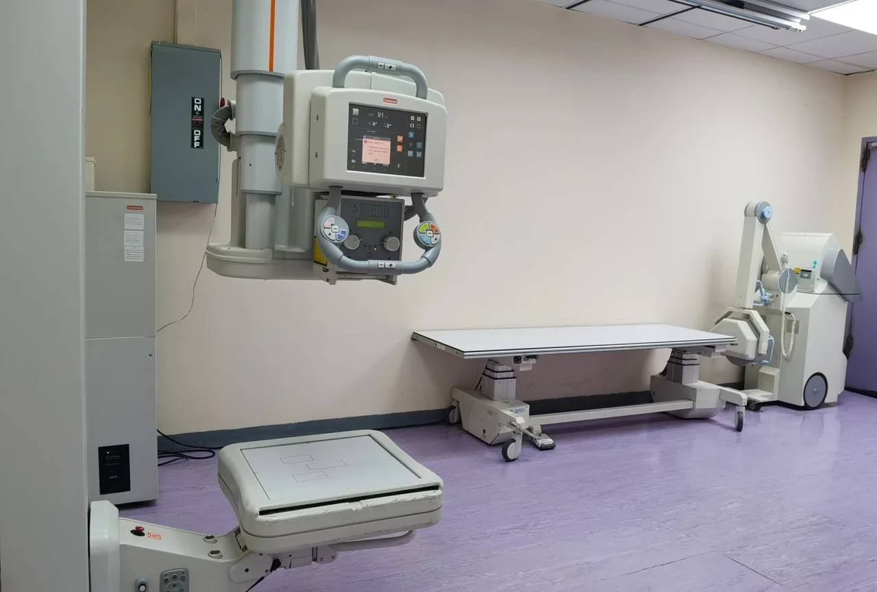
Doctors reach for fluoroscopy when motion or timing matters. Common reasons include:
- Watching how you swallow and how food or liquid moves through the esophagus, stomach and small intestine.
- Guiding a needle, catheter, stent or feeding tube precisely to the right spot inside the body.
- Injecting contrast into a joint, the spine or the urinary tract to study its shape and function.
- Supporting orthopedic, cardiac and vascular procedures where a surgeon needs a live view during the operation.
Because fluoroscopy is often part of a larger diagnostic plan, it may be ordered alongside other studies. Your referring doctor decides whether fluoroscopy, a CT scan, or another test answers the clinical question best.
How a fluoroscopy machine works
The core of the system is an X-ray tube on one side and an image detector, historically an image intensifier, on the other. When the X-ray beam passes through the body, dense tissue such as bone absorbs more of it and appears lighter, while air and soft tissue let more through and appear darker. A camera captures this pattern many times per second, producing a continuous stream of images on a screen.
Modern equipment usually delivers the radiation in short pulses rather than one uninterrupted beam. Pulsed fluoroscopy still looks like smooth motion to the human eye but uses less radiation overall, which is an important safety refinement.
Contrast makes the invisible visible
Soft tissues do not naturally stand out on X-rays. That is why many fluoroscopy exams use a contrast agent. Barium, swallowed or given as an enema, coats the digestive tract. Iodine-based contrast can be injected into blood vessels or joints. The contrast blocks X-rays, so the structure it fills shows up as a bright, well-defined outline you can watch in motion.
Common fluoroscopy procedures
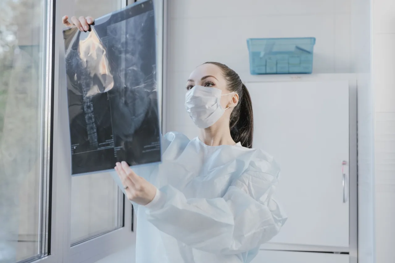
Fluoroscopy underlies a wide family of exams and interventions, including:
- Barium swallow and upper GI series: studying the throat, esophagus, stomach and small bowel as you swallow barium.
- Barium enema: outlining the large intestine.
- Voiding cystourethrogram: watching the bladder fill and empty.
- Arthrogram: injecting contrast into a joint before detailed imaging.
- Angiography and catheter placement: guiding wires and catheters through blood vessels.
- Orthopedic guidance: helping align and fix fractures during surgery.
Some of these overlap with dedicated X-ray studies. For example, if your symptoms point to the spine, your clinician might order a fluoroscopically guided injection or a plain film such as an L-spine X-ray, depending on what they need to see.
How to prepare for a fluoroscopy exam
Preparation depends heavily on which exam you are having, so always follow the specific instructions your clinic gives you. General points often include:
- You may be asked not to eat or drink for several hours before a digestive-tract study.
- You might need to stop certain medications or adjust the timing of doses; confirm this with the ordering clinician.
- Wear comfortable clothing and expect to change into a gown so that zippers, buttons and jewelry do not obscure the images.
- Tell the team if you are or might be pregnant, if you have had a reaction to contrast before, or if you have kidney problems.
What to expect during and after
During the exam you will lie, sit or stand on or beside a movable table. If contrast is used, you will swallow it, receive an enema, or have it injected, depending on the study. The radiologist or technologist will ask you to hold still, change position, or hold your breath at certain moments so the moving images are clear. Most diagnostic fluoroscopy exams take between roughly 15 and 60 minutes.
Afterward, many people return to normal activity right away. If you were given barium, you may be advised to drink extra fluids to help clear it, and your stool may look pale for a day or two. If you had an injection or an interventional procedure, the team will give you specific aftercare and warning signs to watch for.
Radiation safety and risks
Because fluoroscopy delivers X-rays over a longer period than a single radiograph, the radiation dose can be higher, and it varies a lot depending on how long the beam is on and how complex the procedure is. Imaging teams manage this with the ALARA principle, keeping dose As Low As Reasonably Achievable through pulsed beams, tight collimation and careful technique.
For the great majority of patients, the benefit of an accurate diagnosis or a successful guided procedure clearly outweighs the small radiation risk. Extended, complex interventional procedures carry a rare risk of skin effects, and pregnancy calls for extra caution. These trade-offs are exactly the kind of thing to raise with your referring doctor before the exam.
General information, not medical advice
This article is educational and does not diagnose any condition or replace professional care. It cannot tell you what your symptoms or images mean for you. Always discuss your situation, your results and any concerns with your referring doctor, radiologist or another qualified healthcare provider.
Understanding your results
A radiologist, a doctor who specializes in interpreting medical images, reviews the recorded fluoroscopy images and writes a report. That report goes to the clinician who ordered the test, who then discusses the findings with you in the context of your full medical picture. You can read more about what these specialists do in our guide to radiology and how results are read.
Try not to draw conclusions from anything you glimpse on the monitor during the exam. Live images can look dramatic and are easy to misread without training. Wait for the formal report and your clinician's explanation, and bring a written list of questions to that conversation. If you want help choosing where to have imaging done, our directory-style guide on finding and comparing radiology clinics walks through what to look for.
Frequently asked questions
Is fluoroscopy the same as a regular X-ray?
No. A regular X-ray captures a single still image, while fluoroscopy produces a continuous stream of moving images so the team can watch anatomy and function in real time. Fluoroscopy usually runs the beam for longer, so it can involve a higher radiation dose than a single radiograph.
Does fluoroscopy hurt?
The imaging itself is painless. Any discomfort usually comes from the associated procedure, such as swallowing barium, receiving an enema, holding an awkward position, or having a needle or catheter placed. Tell your team if you are uncomfortable, and they can often help.
How much radiation does a fluoroscopy exam involve?
The dose varies widely depending on the exam and how long the beam is used. Because fluoroscopy can use more radiation than a plain X-ray, imaging teams follow the ALARA principle to keep exposure as low as reasonably achievable. For most patients the diagnostic benefit outweighs the small risk, and your doctor can discuss your individual situation.
Do I need contrast for every fluoroscopy exam?
Not always, but many fluoroscopy studies use a contrast agent such as barium or iodine so that soft structures like the digestive tract or blood vessels become visible. Whether you need contrast depends on the specific exam your doctor ordered.
Who interprets my fluoroscopy results?
A radiologist reviews the images and writes a report, which is sent to the doctor who ordered the test. That clinician then explains what the findings mean for you. This article cannot interpret your results, so please discuss them with your healthcare provider.
More guides on MRI, CT & X-Ray
Have a question or a topic to suggest?
We read every message. Tell us what you would like to see next.
You may also like
Hip X-Ray
A hip X-ray is a quick, low-dose imaging test that shows the bones of the hip joint and upper thigh. Doctors use it to look for fractures, arthritis
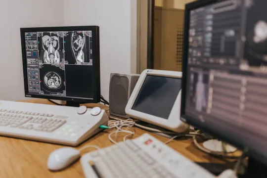
Cect Scan
CECT stands for contrast-enhanced computed tomography, a CT scan performed with a contrast dye to make organs, blood vessels and abnormal areas stand out more clearly.
Low Dose CT Scan for Lung Cancer
A low-dose CT scan for lung cancer, often shortened to LDCT, is a specialized chest scan used to screen people at high risk for lung cancer before symptoms appear.