L Spine X-Ray
An L spine x-ray, short for lumbar spine x-ray, is a quick imaging test that takes pictures of the five bones in your lower back.
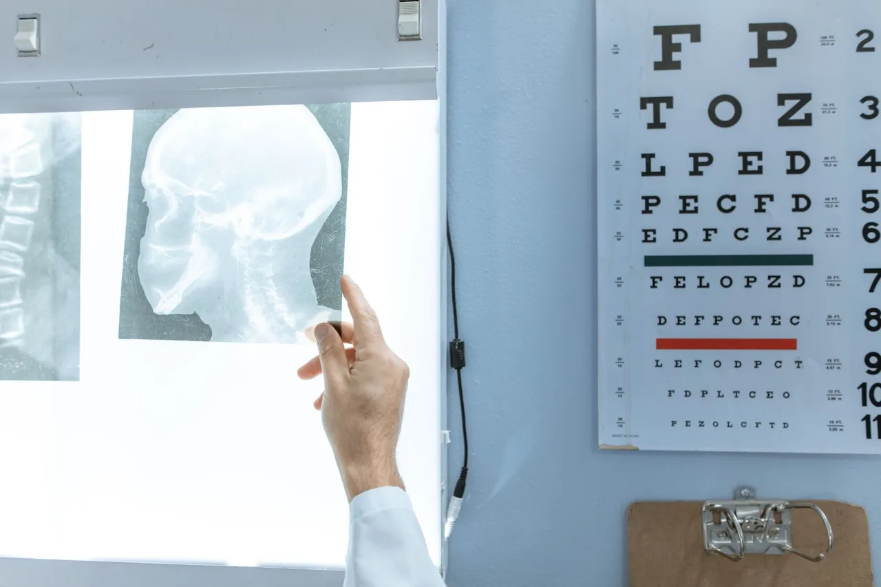
An L spine x-ray, short for lumbar spine x-ray, is a quick imaging test that takes pictures of the five bones in your lower back. Doctors use it to look for fractures, alignment problems, arthritis, and other bone changes behind back pain. This guide explains why an L spine x-ray is ordered, how the procedure works, the small radiation involved, and why only a doctor can tell you what your particular result means.
The short version
- The lumbar spine is the lower part of the back, made up of five vertebrae labelled L1 to L5. An L spine x-ray images these bones.
- It is fast and painless, usually taking only a few minutes, and uses a small dose of radiation.
- X-rays show bone well but reveal little about discs, nerves, and other soft tissue, which is why an MRI is sometimes needed instead.
- A radiologist interprets the images and reports to your doctor. Do not try to read a lumbar spine x-ray on your own.
What an L spine x-ray is
The lumbar spine is the lower section of your backbone. It carries much of your body weight and is made up of five vertebrae, numbered from L1 at the top to L5 near the pelvis. An L spine x-ray passes a small, controlled beam of radiation through this region to create an image on which bone appears white, soft tissue appears grey, and air appears black.
According to RadiologyInfo.org, bone x-rays like this one are the fastest and easiest way for a doctor to view the skeleton and are widely used to diagnose fractures, dislocations, arthritis, and abnormal bone changes. For a wider picture of how this test sits among other scans, see our overview of radiology.
Why a doctor orders a lumbar spine x-ray
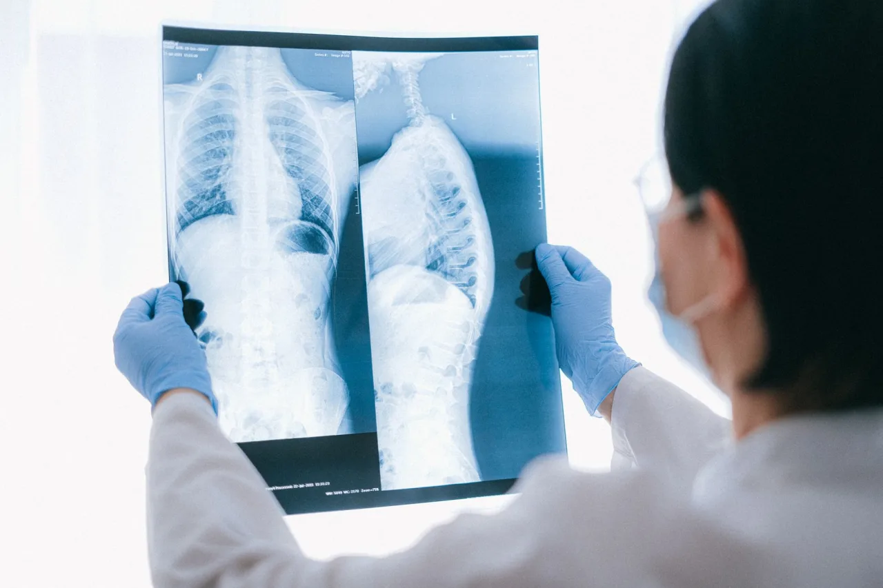
Lower back pain is extremely common, and most cases settle without any imaging at all. When a doctor does request an L spine x-ray, it is usually to check the bones for a specific concern rather than to explain everyday muscular back pain. Common reasons include:
- Assessing a possible fracture after a fall, accident, or injury.
- Looking at spinal alignment, including curvature such as scoliosis or a slipped vertebra.
- Investigating persistent back pain that has warning features or has not improved.
- Checking for arthritis and age-related wear in the joints of the spine.
- Following up on a known bone condition over time.
Because an x-ray shows bone rather than the discs and nerves themselves, a normal L spine x-ray does not rule out every cause of back pain. If your doctor suspects a disc, nerve, or spinal cord problem, they may order an MRI scan instead, because it shows soft tissue far more clearly.
How the procedure works, step by step
An L spine x-ray is usually quick and straightforward. The whole appointment often takes only a few minutes.
- You may be asked to change into a gown and to remove jewellery, belts, or anything metal near your lower back.
- A radiographer positions you against or on top of the imaging plate, often standing, lying on your back, and lying on your side.
- You hold still, and are sometimes asked to hold your breath for a second, while each image is taken.
- The radiographer steps behind a screen to take the picture, then repositions you for the next view.
Several images are usually taken so the vertebrae can be seen from different angles. As the NHS explains, the x-ray itself is painless, although lying still on a firm table can be briefly uncomfortable if your back is already sore. You will not feel the radiation, and none stays in your body afterwards.
Tip: Tell the radiographer if you are or might be pregnant before any x-ray. The NHS notes that scans are generally avoided during pregnancy where possible, and staff can discuss alternatives or extra precautions with you.
Radiation and safety
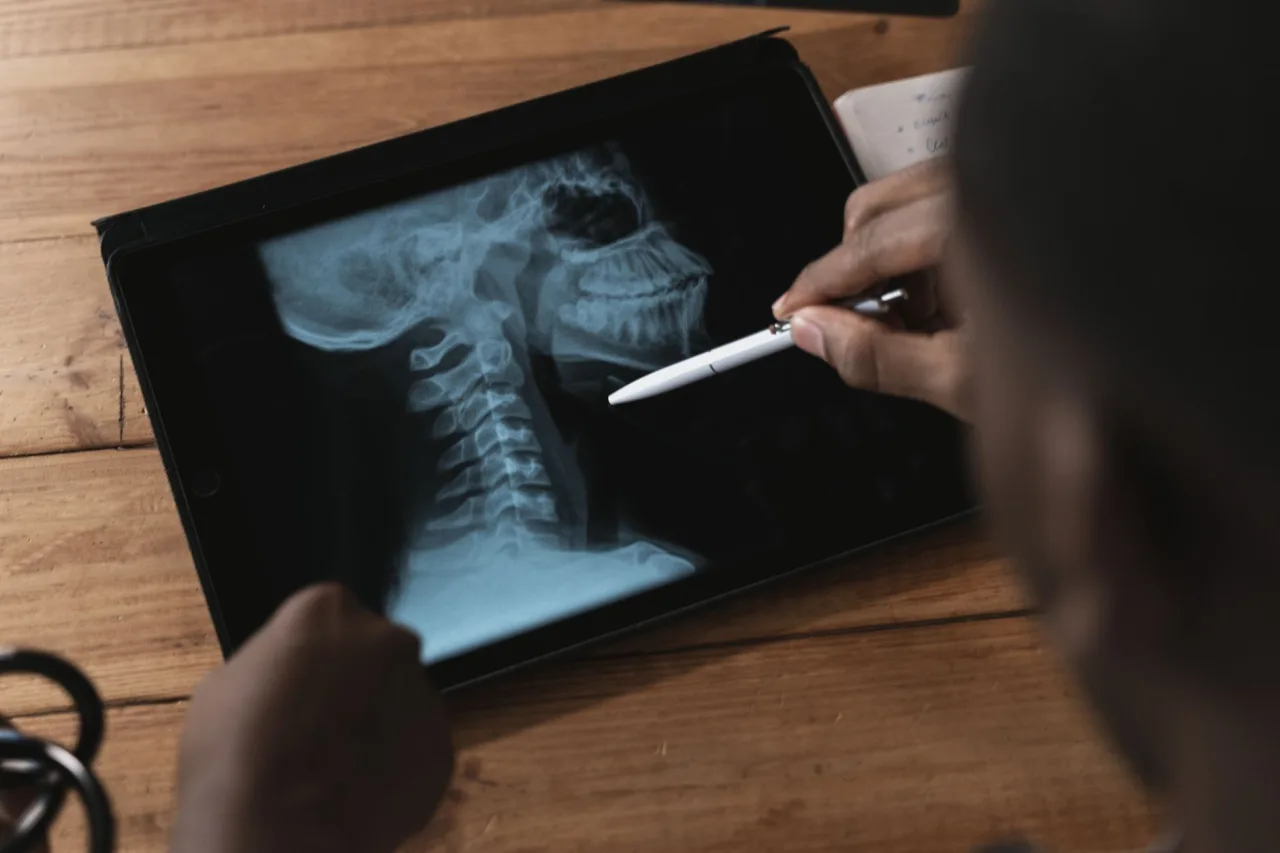
An L spine x-ray uses ionising radiation, but only a small amount. RadiologyInfo.org describes bone x-rays as using a very low dose, and notes that x-rays usually have no side effects in the typical diagnostic range. Because the lower spine sits near the abdomen and reproductive organs, the dose for a lumbar study is a little higher than for, say, a hand or foot, which is one reason doctors order it only when it will genuinely help.
The important point is proportion. For a person with a suspected fracture or a spine that needs assessing, the information from the image usually far outweighs the small radiation risk. Facilities follow the ALARA principle, meaning as low as reasonably achievable, using the smallest dose that still produces a clear picture. If you have had several spine x-rays already, it is reasonable to ask your doctor whether another is necessary.
What your results mean
After the images are taken, a radiologist examines them and writes a report for the doctor who requested the test. That doctor then explains the findings alongside your symptoms, examination, and history. A radiology report often lists observations such as disc space narrowing, bone spurs, or mild alignment changes, and these are common with age and do not always cause pain.
This is exactly why you should not interpret an L spine x-ray yourself. The same finding can be significant in one person and completely harmless in another, and reports are written in cautious, technical language meant for clinicians. Terms that sound alarming, such as degeneration or wear, are frequently normal for your age and unrelated to the reason you had the scan.
This is general information, not medical advice. A lumbar spine x-ray cannot be interpreted in isolation, and the same image can mean different things for different people. Do not draw conclusions from your images or an online report. Always discuss the results with the doctor who ordered the scan or a specialist they refer you to.
How an L spine x-ray compares with other tests
An x-ray is often the first-line test for the bony spine because it is fast, widely available, and low cost. It is excellent for showing fractures, alignment, and arthritis in the vertebrae. What it cannot show well are the intervertebral discs, the spinal cord, and the nerves that leave the spine, all of which are soft tissue.
When the suspected problem involves those structures, such as a herniated disc pressing on a nerve, an MRI is usually the better test because it images soft tissue in detail without using radiation. A CT scan may be used for complex fractures or detailed bone assessment. Your doctor chooses the test that answers the specific question, and a lumbar x-ray is frequently the sensible starting point. If you are comparing a lumbar with a neck study, our guide to the C spine x-ray covers the cervical region, and the hip x-ray guide covers a nearby joint that can also refer pain to the back.
Cost and where to have an L spine x-ray
What an L spine x-ray costs varies widely by country, provider, and whether it is covered by insurance or a public health system. Plain x-rays are generally among the cheaper imaging tests, but the same study can differ noticeably between facilities, so it is always worth confirming both the price and your coverage with the clinic and your insurer before booking.
Choosing an accredited, well-run clinic matters more than choosing the closest one. Our guide to finding a good x-ray clinic near you explains what to look for, and the technologists who operate these machines follow a defined training and certification path, described in our look at the x-ray technologist role.
Frequently asked questions
What does L spine x-ray stand for?
The L stands for lumbar, meaning the lower back. An L spine x-ray images the five lumbar vertebrae, labelled L1 to L5, which sit between the chest region and the pelvis. It is simply a plain x-ray focused on this part of the spine.
Does an L spine x-ray hurt?
The x-ray itself is painless, and you will not feel the radiation. The only discomfort some people notice is from lying still on a firm table or moving into different positions when the back is already sore. Each position is held only for a few seconds.
Can a lumbar spine x-ray show a slipped disc?
Not directly. An x-ray shows the bones and can hint at a problem through changes such as narrowed disc spaces, but it cannot show the disc itself or a nerve being pressed. If a slipped disc is suspected, a doctor usually orders an MRI, which images soft tissue clearly.
How long does the appointment take?
The x-ray itself usually takes only a few minutes, though the full appointment is a little longer to allow for changing, positioning, and taking several views. Most people are in and out quickly, and there is no recovery time afterwards.
Who tells me the results of my L spine x-ray?
A radiologist interprets the images and sends a report to the doctor who ordered the scan. That doctor explains what the findings mean for you in the context of your symptoms. You should not rely on reading the images or the report yourself, because interpretation depends on your full clinical picture.
More guides on MRI, CT & X-Ray
Have a question or a topic to suggest?
We read every message. Tell us what you would like to see next.
You may also like

Foot X-Ray Normal
A foot x-ray is a quick, painless test that shows the many small bones and joints of the foot. If you have searched for foot x-ray normal, you are probably trying to understand what a normal result actually means
CT Scan Abdomen and Pelvis
A CT scan of the abdomen and pelvis uses x-rays and a computer to build detailed cross-sectional pictures of the organs in your belly and lower trunk.
MRI Center
An MRI center is a facility, either inside a hospital or as a standalone clinic, that performs magnetic resonance imaging. If your doctor has referred you for an MRI, knowing what happens at one of these centers