Mobile Imaging
Mobile imaging means bringing the scanner to the patient instead of the patient to the scanner.
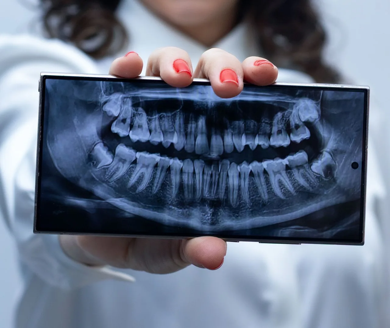
Mobile imaging means bringing the scanner to the patient instead of the patient to the scanner. Portable X-ray units, handheld ultrasound probes and truck mounted MRI or CT scanners let care teams take pictures at the bedside, in a care home, or in a rural community. This guide explains what mobile imaging is, why it is used, how a mobile scan works and how to read the report that follows.
The short version
- Mobile imaging delivers scans such as X-ray, ultrasound and sometimes CT or MRI at a location away from a hospital radiology department.
- It is used for patients who cannot travel easily, for rural and community access, and for fast answers at the bedside.
- Image quality and available scan types can differ from a fixed department, so the setting is chosen to fit the clinical question.
- A radiologist still interprets the images, and only your doctor can tell you what the findings mean for you.
What mobile imaging is
Mobile imaging is a broad term for medical scans performed with equipment that travels to the patient rather than sitting permanently in a hospital. The category covers several very different setups. At the smallest end are handheld and cart based ultrasound machines that a technologist wheels to a hospital bed. In the middle sit portable X-ray units used on wards and in care homes. At the largest end are full CT and MRI scanners built into trailers or trucks that park outside a clinic and connect to power.
The common thread is convenience and access, not a different kind of physics. A mobile X-ray still uses the same ionizing radiation as a department X-ray, and a portable ultrasound still uses the same high frequency sound waves as a large console. If you want the underlying principles first, our overview of how medical imaging works and our guide to what an ultrasound is both explain the modalities that mobile services deliver.
Common types of mobile imaging
Different mobile setups suit different questions. The most common include:
- Portable X-ray: a wheeled unit brought to a bed or a care home room, often to check the chest, an abdomen or a possible fracture when a patient cannot be moved easily.
- Point of care and portable ultrasound: small machines used at the bedside, in clinics and by community teams. See our explainer on the portable USG machine for how these work.
- Mobile CT and MRI: full scanners in trailers that visit hospitals during upgrades, or serve areas without a fixed unit. These require careful siting, power and shielding.
- Mobile mammography: screening buses that bring breast imaging to workplaces and underserved communities.
Why a mobile scan is used
Clinicians and services choose mobile imaging for practical reasons rather than because it is a superior test. Typical drivers include:
- Patients who are too unwell, frail or immobile to travel to a department, including hospital inpatients and care home residents.
- Access for rural and remote communities that are far from a fixed imaging center.
- Speed at the bedside, for example a portable chest X-ray or a focused ultrasound to guide urgent decisions.
- Screening outreach, such as a mammography bus visiting a workplace or town.
- Covering a hospital scanner that is temporarily out of service for maintenance or replacement.
For services that need equipment they can move between sites, buying decisions matter. Our buyer's guide to portable ultrasound machines looks at what clinics weigh up when choosing kit for mobile work.
How a mobile scan works
The scan itself follows the same steps as its department equivalent. A mobile X-ray technologist positions a small detector behind the body part, aligns the portable tube head and takes the exposure, shielding nearby people. A portable ultrasound examiner applies gel and moves a probe over the skin, watching live images on the screen. A mobile CT or MRI works like a fixed unit inside the trailer, with the same positioning, breath holds and, where needed, contrast.
Once acquired, images are usually sent electronically to a radiologist through a secure network, a workflow that is standard across modern radiology. That means the person taking the pictures and the person interpreting them are often in different places, which is normal and does not reduce the quality of the reading.
Tip: Ask the mobile service how your images will reach a radiologist and how the report will get back to your referring doctor. A clear reporting pathway matters more than where the machine happens to be parked.
Benefits and limits
The main benefit of mobile imaging is access. It reaches people who would otherwise struggle to be scanned, brings screening closer to communities, and can deliver fast answers at the bedside. For a frail patient, avoiding an ambulance transfer is a real safety and comfort gain.
There are trade offs. Portable X-ray images can be technically harder than department films because positioning is limited at a bedside. Point of care ultrasound is focused on a specific question rather than a full survey. Mobile CT and MRI need suitable siting, power and safety zones. Not every scan type is available in every mobile setting, so the service and the referring clinician match the setup to what needs answering. When a fuller study is needed, you may still be asked to visit a fixed department for something like a detailed kidney sonogram or a cross sectional scan.
Safety and radiation
Safety depends on the modality, not on whether it is mobile. Ultrasound and MRI use no ionizing radiation. Ultrasound uses sound waves and, according to RadiologyInfo.org, standard diagnostic ultrasound has no known harmful effects on humans. MRI uses a strong magnetic field and radio waves, with the important caveat that all metal must be declared and removed because the magnet is always on. X-ray, CT and mammography use ionizing radiation in carefully controlled, low doses, and the small risk is weighed against the benefit of the information. Mobile X-ray teams still follow shielding and distance rules to protect patients, staff and bystanders.
Understanding your results
A mobile scan produces the same kind of images as a department scan, and a radiologist reviews them and writes a report for the doctor who referred you. You will usually not get a full result on the spot, because the person operating a mobile unit is often a technologist rather than the radiologist who interprets the study.
Reports are descriptive. They record what the images show, for example clear lungs, a possible fracture, fluid, or an area that needs another test. These observations are not a diagnosis by themselves. Their meaning depends on your symptoms, your history and other tests, which is why the same finding can matter very differently for two people.
Important: This article is general educational information, not medical advice, and it cannot tell you what your own scan means. Please discuss your results and any next steps with your referring doctor or a radiologist.
How to choose a mobile imaging service
If you or a family member are arranging a mobile scan, focus on quality and the reporting pathway rather than convenience alone. Sensible checks include:
- Confirm the service is properly accredited and that scans are performed by qualified technologists.
- Ask who interprets the images and how quickly a radiologist report reaches your doctor.
- Check that images are stored so future scans can be compared.
- Clarify the cost, what it includes and how insurance is handled before booking.
Costs vary widely by country, provider and whether you pay privately or use insurance, so confirm the fee and your coverage in advance. If you are comparing options, our directory guide to finding X-ray services near you covers the same quality questions you can ask any mobile provider.
Frequently asked questions
What does mobile imaging mean?
Mobile imaging means a medical scan performed with equipment that travels to the patient rather than being fixed in a hospital. It includes portable X-ray and ultrasound at the bedside and truck mounted CT or MRI units, used to reach people who cannot easily travel to a department.
Is a mobile scan as good as a hospital scan?
It uses the same underlying technology and is interpreted by a radiologist in the same way. For many questions it is fully adequate. Some setups are more focused or face harder positioning than a fixed department, so the service and your doctor match the mobile option to the clinical question.
Does mobile imaging use radiation?
It depends on the type. Mobile ultrasound and MRI use no ionizing radiation. Mobile X-ray, CT and mammography use controlled low doses of ionizing radiation, with shielding and distance rules to protect patients and staff, just like their department equivalents.
Who performs and reads a mobile scan?
A qualified technologist or sonographer usually performs the scan, and a radiologist reviews the images and writes the report. The two are often in different locations, which is normal and does not reduce quality.
When will I get my mobile imaging results?
You will rarely get a full result on the spot. The images are sent to a radiologist who interprets them and reports to your referring doctor, often within a few days, and your doctor then explains what the findings mean for you.
Sources and further reading:
- General Ultrasound Imaging, RadiologyInfo.org for how ultrasound works and its safety profile.
- Ultrasound scan, NHS for ultrasound types and why it uses no radiation.
- Magnetic Resonance Imaging (MRI), RadiologyInfo.org for how MRI works and its metal safety rules.
More guides on Ultrasound & Radiology (General)
Have a question or a topic to suggest?
We read every message. Tell us what you would like to see next.
You may also like

Wave Imaging
If you searched for "wave imaging," you may be looking for one of two things: a specific imaging center that uses the name, or the broader idea that most medical scans are built on waves of one kind or another.

Kidney Sonogram
A kidney sonogram, also called a renal ultrasound, is a safe, radiation free scan that shows the size, shape and structure of your kidneys and nearby urinary tract.

Village Imaging
If you have searched for "village imaging" you are probably looking for a local, community based imaging center rather than a single national brand.