KUB X-Ray: Uses, Procedure & Results
What a KUB X-ray looks at, how the appointment works, the radiation involved, and how doctors interpret the results.
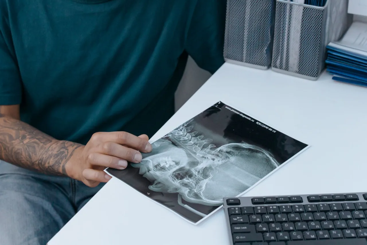
A KUB X-ray is one of the quickest, lowest-cost imaging tests in medicine: a single flat picture of your kidneys, ureters and bladder that a doctor can read within minutes. If your clinician has ordered one, or you have seen the term "KUB" on a request slip, this guide explains what the scan looks at, how the appointment works, and how radiologists make sense of the image.
The short version
- KUB stands for Kidneys, Ureters and Bladder. It is a plain abdominal X-ray focused on the urinary tract.
- It uses a very small dose of ionizing radiation and takes only about 10 to 15 minutes, with no needles and no special recovery.
- It is often the first step to look for kidney stones, bowel gas patterns, blockages or swallowed objects, but it is not a detailed test for soft tissue.
- Only a qualified doctor can interpret your images alongside your symptoms. Never self-diagnose from a report line.
What a KUB X-ray actually is
KUB is short for Kidneys, Ureters and Bladder. It is a standard abdominal X-ray, taken from the front, that is centered so the whole urinary tract sits within the picture. Because it is a single exposure rather than a series, it is fast, widely available and inexpensive compared with cross-sectional scans.
According to RadiologyInfo.org, an abdominal X-ray evaluates internal organs including the stomach, liver, intestines and spleen, and when it is aimed at the kidneys, ureters and bladder it is described as a KUB study. The same image therefore doubles as a general survey of the abdomen and a focused look at the urinary system. For a broader look at how these films are used, see our guide to the abdominal X-ray.
Why a doctor might order one
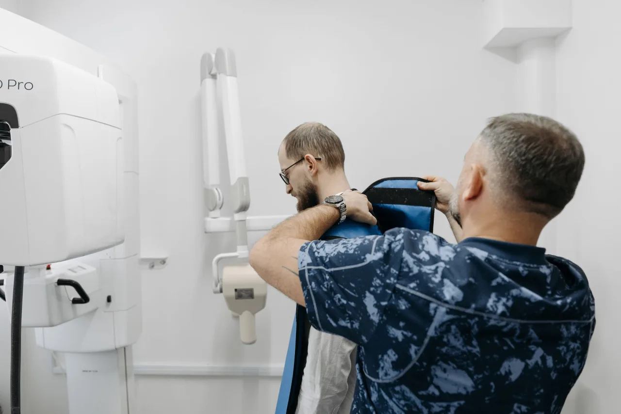
A KUB is usually requested when a clinician wants a fast, first-pass answer rather than fine anatomical detail. Common reasons include:
- Suspected kidney or bladder stones, especially stones dense enough to show on plain film.
- Assessing bowel gas patterns when a blockage (obstruction) or a perforation is a concern.
- Checking the position of a device such as a ureteric stent or a catheter tip.
- Looking for a swallowed foreign object.
- Following up a known finding over time without repeated higher-dose scans.
MedlinePlus notes that an abdominal film shows organs and structures such as the liver, spleen, stomach, intestines and lower spine, and that when it is focused on the urinary structures it is called a KUB. It is a screening and triage tool: if it raises a question, your doctor may order a more detailed study next.
How it works
X-rays are a form of energy that passes through the body at different rates depending on tissue density. Dense structures such as bone and many stones absorb more of the beam and appear white, soft tissues appear in shades of gray, and gas or air appears black. The detector behind you records the pattern, producing the familiar image a radiologist reviews.
Because a KUB is a two-dimensional projection, overlapping structures can hide or mimic each other. That is a key reason it is often paired with, or followed by, ultrasound or CT when more certainty is needed. If your care team is weighing options, our overviews of the CT scan of the abdomen and pelvis and of ultrasound explain how those tests add different information.
How to prepare
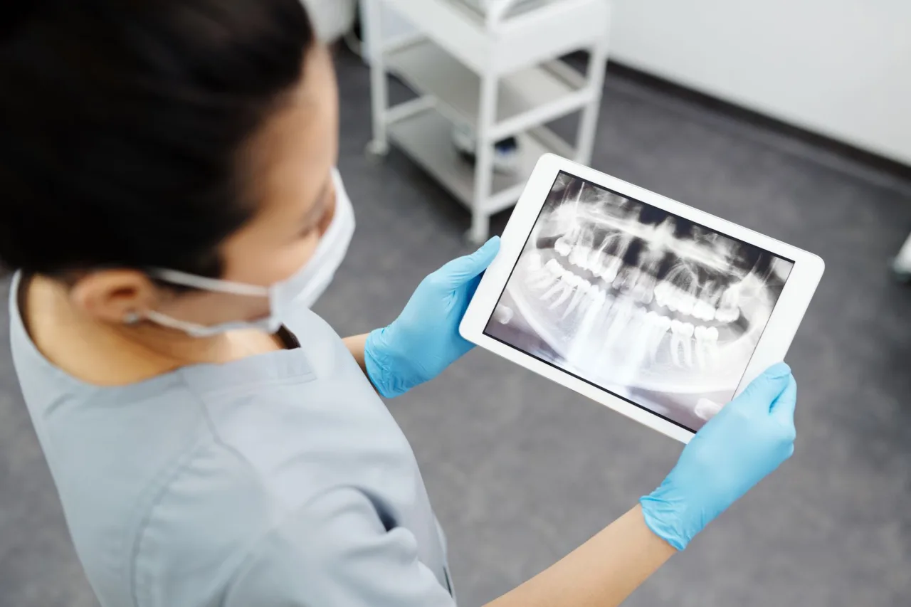
Preparation for a KUB is minimal. In most cases you can eat and drink normally unless your clinic gives you specific instructions. RadiologyInfo.org advises removing jewelry and metal objects, telling the technologist about any recent barium studies or bismuth-containing medicines (which can obscure the picture), and informing staff if you are or might be pregnant, or if you use an IUD.
Before you go
- Wear comfortable clothing without metal fasteners; you may be given a gown.
- Tell the team if you could be pregnant, so alternatives or shielding can be considered.
- Bring your request form and a list of recent imaging so studies are not needlessly repeated.
What happens during the scan
The appointment itself is short. A radiographer positions you, usually lying on your back on the X-ray table, and may ask for additional views on your side or while standing to see how gas and fluid shift. MedlinePlus notes that a lead apron or shield may be used to limit exposure to areas that do not need imaging. You will be asked to hold still, and sometimes to hold your breath for a moment, while the exposure is taken. There is no needle and no discomfort from the imaging itself, and RadiologyInfo.org notes the exam is typically completed within about 15 minutes.
Radiation and safety
A KUB uses ionizing radiation, but only a very small dose. RadiologyInfo.org describes the exposure as very small and states that the benefit of an accurate diagnosis far outweighs the associated risk. MedlinePlus adds that X-ray equipment is monitored and regulated to deliver the minimum radiation needed to make the image.
The main precaution is pregnancy. Because a developing baby is more sensitive to radiation, plain abdominal X-rays are generally avoided during pregnancy unless clearly necessary, and ultrasound (which uses no radiation) is often preferred. Always tell your care team if you are pregnant or think you might be, and ask any questions before the scan.
Important: this is general information, not medical advice. The details here describe how KUB X-rays generally work. They cannot tell you what your own scan means. Only your referring clinician or a radiologist, who can see your images and your full history, can interpret your results and advise on next steps. If you have new, severe or worsening symptoms, contact a healthcare professional promptly.
What the results can and cannot show
A KUB is good at revealing dense or gas-related findings: many stones, abnormal gas patterns that suggest an obstruction or perforation, the outline of enlarged organs, and the position of devices or swallowed objects. RadiologyInfo.org lists blockages, perforations, foreign objects and calcified stones among the things it can detect, which makes it valuable in urgent settings.
Its limits matter just as much. A plain film does not reliably show soft-tissue detail, small or non-calcified stones, early tumors or the inside of organs. A normal KUB does not rule out disease, and a shadow on the film is not a diagnosis. That is why an abnormal or uncertain KUB is frequently followed by ultrasound, CT or another targeted study.
What happens after
A radiologist reviews the images and sends a written report to the doctor who ordered the test. You can usually return to normal activity immediately. Your clinician will combine the report with your symptoms, examination and any blood or urine tests before deciding what, if anything, comes next. If a follow-up scan is recommended, you may find it helpful to understand costs in advance; our guide to the price of a CT scan covers how imaging is typically billed.
Frequently asked questions
Does a KUB X-ray hurt?
No. The imaging itself is painless. You simply lie still on the table while the pictures are taken. There are no needles for a standard KUB, and there is nothing to recover from afterward.
How long does a KUB X-ray take?
The scan is quick, usually around 10 to 15 minutes including positioning and any extra views. The actual exposures last only a fraction of a second each. You can normally go about your day immediately afterward.
Is the radiation dangerous?
A KUB uses only a very small dose of ionizing radiation, and authorities such as RadiologyInfo.org state that the diagnostic benefit generally outweighs the small risk. The main exception is pregnancy, when radiation-free options like ultrasound are usually preferred. Tell your team if you could be pregnant.
Do I need to fast or prepare?
Usually not. Most people can eat and drink normally before a KUB unless the clinic advises otherwise. You will be asked to remove metal objects and jewelry, and to mention any recent barium tests, which can obscure the image.
Can a KUB detect kidney stones?
It can show many stones that are dense enough to appear on a plain film, which is one of its most common uses. However, it can miss smaller or non-calcified stones, so a normal KUB does not fully rule them out. A doctor may add ultrasound or CT if stones are strongly suspected.
What is the difference between a KUB and a CT scan?
A KUB is a single, low-dose, two-dimensional X-ray that is fast and inexpensive. A CT scan builds detailed cross-sectional images and shows far more soft-tissue detail, but uses more radiation and costs more. Your clinician chooses based on what question needs answering.
Sources: RadiologyInfo.org, Abdominal X-ray (KUB); MedlinePlus, Abdominal X-ray; NHS, X-ray.
More guides on MRI, CT & X-Ray
Have a question or a topic to suggest?
We read every message. Tell us what you would like to see next.
You may also like

X-Ray Scan
An X-ray scan is one of the fastest and most common medical imaging tests, often done in minutes to check bones, the chest and more. This guide explains what an X-ray exam involves, why doctors order it
CT Scan Abdomen and Pelvis
A CT scan of the abdomen and pelvis uses x-rays and a computer to build detailed cross-sectional pictures of the organs in your belly and lower trunk.
Ultrasound: Uses, Procedure & Results
An ultrasound uses sound waves, not radiation, to image soft tissue in real time. This guide covers why scans are ordered, how they work, how to prepare, and how results are interpreted, with a reminder to discuss findings with your doctor.