8 Week Ultrasound: Uses & Results
The early scan explained: why it is done, why it is often internal, what it shows and how to read the results.
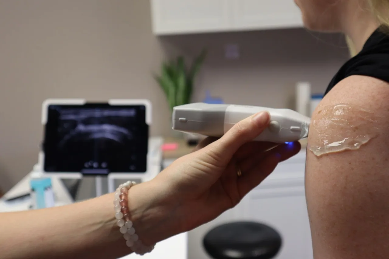
An 8 week ultrasound is one of the earliest scans in pregnancy, often done to confirm a pregnancy is developing in the right place, to check for a heartbeat, and to help work out dates. Unlike the routine dating scan later on, an 8 week sonogram is frequently arranged for a specific reason, and it is commonly performed internally because the baby is still so small. This guide explains why the scan is done, what it involves, and how to think about the results, with the reminder that your own clinician is the one who can interpret them.
The short version
- An 8 week ultrasound is an early scan, often arranged for reassurance, symptoms, or after fertility treatment.
- At this stage it is commonly done as a transvaginal (internal) scan for a clearer view.
- It can confirm the pregnancy's location, check for a heartbeat, and help estimate dates.
- Findings this early can be uncertain, so follow-up scans are sometimes needed and results should be discussed with your clinician.
What an 8 week ultrasound is
An 8 week ultrasound is an early pregnancy scan performed near the start of the first trimester. In many health systems the first routine scan is the later dating scan, so an 8 week scan is often an "early scan" arranged for a particular reason rather than a universal appointment. Its purpose is usually to confirm that a pregnancy is in the uterus, to look for a heartbeat, and to measure the tiny embryo to help estimate gestational age. Confirming the location matters because a pregnancy that develops outside the uterus needs prompt medical attention, and an early scan is one of the tools clinicians use to check this. It is worth setting expectations gently: at 8 weeks the aim is basic confirmation and dating rather than the detailed anatomy assessment that comes much later in pregnancy. For the wider journey, our week-by-week guide to pregnancy sonograms shows how the early scan fits with later ones.
Why an 8 week scan is done

Because it is not always routine, an 8 week ultrasound is often prompted by a specific situation. Common reasons include:
- Reassurance: checking for a heartbeat, sometimes after a previous loss.
- Symptoms: bleeding or pain in early pregnancy, which need prompt assessment.
- Uncertain dates: when the timing of the last period is unknown or irregular.
- After fertility treatment: an early viability scan is common following IVF.
- Checking the location: confirming the pregnancy is in the uterus rather than elsewhere.
Important
Bleeding or significant pain in early pregnancy should always be assessed promptly by a healthcare professional or an early pregnancy unit, not managed by booking a private keepsake scan. If you have these symptoms, contact your midwife, doctor or local early pregnancy service.
How an 8 week ultrasound is done
At 8 weeks the embryo is only around the size of a raspberry, so a scan through the abdomen may not give a clear enough view. For this reason an 8 week scan is often performed transvaginally, using a slim probe gently inserted into the vagina, which sits much closer to the uterus and gives a sharper early image. Our explainer on the transvaginal ultrasound describes exactly what that involves and how it feels. Some clinics will try an abdominal approach first, with a full bladder, and only suggest the internal scan if the view is unclear. If ultrasound is new to you, our overview of how ultrasound works is a helpful primer.
How to prepare for an early scan
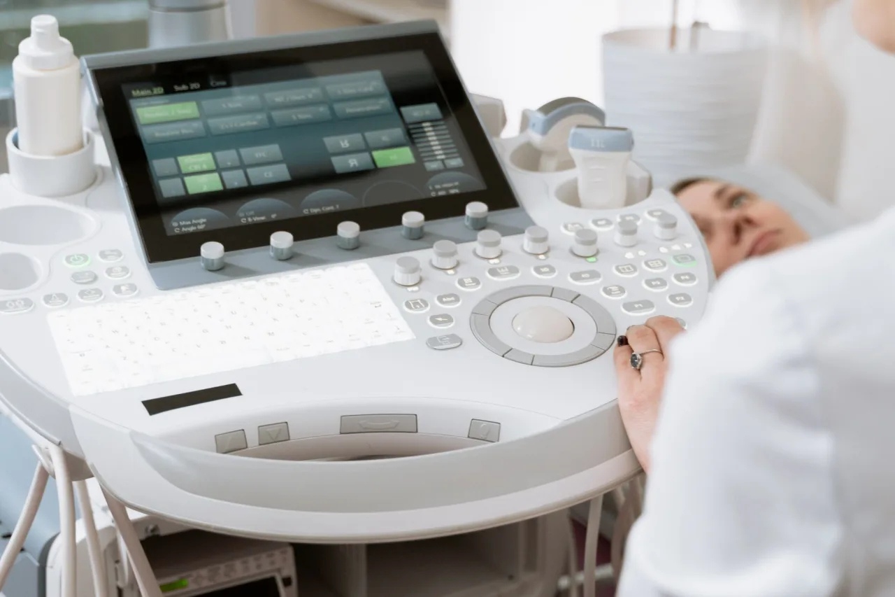
Preparation depends on which approach the clinic expects to use, so it is worth asking when you book. A few general pointers help:
- For a transvaginal scan: you usually need an empty bladder, which is the opposite of the later dating scan, so avoid arriving having deliberately filled up.
- For an abdominal attempt: the clinic may ask for a full bladder to improve the view, so follow whatever instruction you are given rather than guessing.
- Wear comfortable, two-piece clothing so you can undress easily from the waist down if an internal scan is needed.
- Expect a short appointment, though the emotional weight of an early scan can make it feel longer, so allow yourself time afterwards.
You are entitled to ask questions and to have a supporter with you where the clinic allows it. If an internal scan is suggested and you are anxious about it, tell the sonographer, who can explain each step and go at your pace.
What you might see at 8 weeks
By around 8 weeks a scan can often show the pregnancy sac, the embryo, and a heartbeat, and the sonographer may measure the embryo from crown to rump to estimate dates. That said, what is visible varies with exact timing, the equipment, and the position of the uterus. If a scan a little earlier does not yet show everything expected, it does not automatically mean something is wrong, and a repeat scan a week or two later is a common and sensible next step. Try not to read too much into a single very early image. Our guides to the 6 week ultrasound and the 12 week ultrasound explain how the picture changes over just a few weeks.
Is an 8 week ultrasound safe?
Ultrasound uses sound waves rather than radiation, which is a major reason it is used throughout pregnancy. The NHS states there are no known risks to the baby or the mother from having an ultrasound scan, and that scans are painless with no known side effects. RadiologyInfo.org similarly notes that ultrasound has been used in pregnancy for decades with no evidence of harm to the patient, embryo or fetus when used appropriately by trained professionals. Transvaginal scanning in early pregnancy is considered safe when done by a qualified operator.
What the results mean and what comes next
An early scan can be wonderfully reassuring, but it can also be a moment of uncertainty, because findings at 8 weeks sometimes need a follow-up to become clear. One common example is when the dates turn out to be earlier than expected, so a structure that has not appeared yet may simply be a matter of timing rather than a problem. In these situations clinicians often recommend a repeat scan, sometimes alongside blood tests, before drawing any conclusions, precisely because acting on a single ambiguous early image can be misleading. This waiting can be hard, and it is completely reasonable to ask your clinician what the plan is and when you will be seen again.
Whatever the scan shows, the person to interpret it is the clinician caring for you, who can put the images together with your dates, symptoms and any blood tests. Please do not try to reach a conclusion from the images or measurements alone, and be wary of comparing your scan against strangers' stories online, because early pregnancy varies so much from person to person. If you are booking privately for reassurance, choose a reputable, well-run clinic; our guide to finding a good private ultrasound provider explains what to look for.
A note on medical advice
This article is general educational information, not medical advice, and it cannot tell you what your own scan means. For anything specific to your pregnancy, especially if you have bleeding or pain, speak with your midwife, doctor or local early pregnancy service.
Frequently asked questions
Is an 8 week ultrasound done internally or on the tummy?
At 8 weeks the embryo is very small, so the scan is often done transvaginally, with a slim internal probe, to get a clearer view. Some clinics try an abdominal scan first with a full bladder and only switch to an internal scan if the view is not clear enough.
Will I see a heartbeat at 8 weeks?
A heartbeat can often be seen by around 8 weeks, but this varies with exact timing, the equipment and the position of the uterus. If a very early scan does not yet show everything expected, it does not automatically mean something is wrong, and a repeat scan a week or two later is a common next step.
Why would I have a scan this early?
An 8 week scan is often arranged for a specific reason, such as reassurance after a previous loss, bleeding or pain, uncertain dates, or an early viability check after fertility treatment. It may also confirm that the pregnancy is developing in the right place.
Is an early transvaginal scan safe in pregnancy?
Yes. Ultrasound uses sound waves rather than radiation, and the NHS states there are no known risks to the baby or the mother from having an ultrasound scan. Transvaginal scanning in early pregnancy is considered safe when performed by a qualified operator.
What should I do if I have bleeding or pain in early pregnancy?
Bleeding or significant pain in early pregnancy should be assessed promptly by a healthcare professional or an early pregnancy unit, rather than managed by booking a keepsake scan. Contact your midwife, doctor or local early pregnancy service for advice.
More guides on Ultrasound & Radiology (General)
Have a question or a topic to suggest?
We read every message. Tell us what you would like to see next.
You may also like

12 Week Ultrasound: Uses & Results
A guide to the 12 week dating scan: timing, purpose, preparation, what you might see, combined screening and safety, with a reminder to talk to your clinician.

6 Week Ultrasound: What to Expect
A 6 week pregnancy ultrasound checks location, number, and an early heartbeat. Learn what can be seen this early, how the scan is done, and why repeat scans are normal.

Pregnancy Sonogram: Week by Week
A reassuring, week by week look at pregnancy ultrasound scans, including what the dating and anomaly scans check, how to prepare and how results are shared.