USG Abdomen Scan: Uses and Procedure
How the abdominal ultrasound works, how to prepare, and why results belong with your doctor.

A USG abdomen scan, better known as an abdominal ultrasound, uses harmless sound waves to look at the organs inside your belly in real time. It is painless, involves no radiation, and is one of the most common first tests a doctor orders for abdominal pain or abnormal blood results. This guide explains why it is requested, how the scan works, how to prepare, and why only your doctor can tell you what your own results mean.
The short version
- USG stands for ultrasonography. A USG abdomen scan images the liver, gallbladder, kidneys, pancreas, spleen, and major blood vessels.
- It uses sound waves, not radiation, so it is considered very safe and is often the first imaging test ordered.
- You usually need to fast for several hours beforehand so the gallbladder and bowel show up clearly.
- A radiologist interprets the images. Only your doctor can explain what any finding means for you.
What a USG abdomen scan is
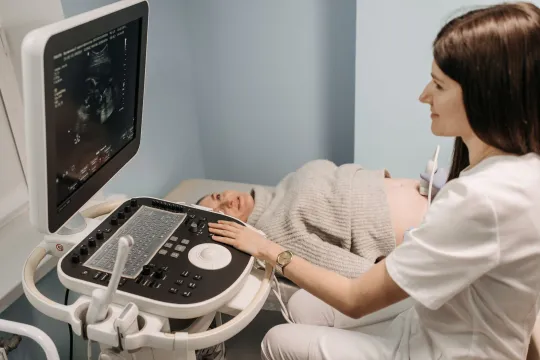
USG is simply an abbreviation of ultrasonography, so a USG abdomen scan and an abdominal ultrasound are the same test under two names. A handheld probe called a transducer is moved over your skin, sending high-frequency sound waves into the body. Those waves bounce off organs and tissues, and the returning echoes are turned into moving pictures on a screen. Because it works in real time, the operator can watch organs move, check blood flow, and measure structures as the exam happens.
According to RadiologyInfo.org, an abdominal ultrasound uses a small probe and gel placed directly on the skin, and it is extremely safe because it does not use radiation. That safety is a big reason doctors reach for it first. For a wider view of how ultrasound fits among all the imaging tests, see our overview of what an ultrasound is and how it works.
Why a doctor orders one

A USG abdomen scan is a versatile diagnostic tool. Your doctor may request it to investigate symptoms, to follow up an abnormal blood test, or to keep an eye on a known condition. Common reasons include:
- Unexplained abdominal pain, bloating, or tenderness.
- Suspected gallstones or gallbladder inflammation.
- Abnormal liver blood tests, or checking for a fatty or enlarged liver.
- Kidney problems, including stones, cysts, or blockage.
- Investigating an aortic aneurysm or assessing the spleen and pancreas.
- Guiding a needle safely during a biopsy or fluid drainage.
Because it is quick, painless, and radiation-free, an abdominal ultrasound is frequently the starting point. If it raises further questions, your doctor may then order a more detailed test such as a CT scan or an MRI. If the concern is focused on one organ, you may be sent for a targeted study such as a liver ultrasound.
How to prepare
Preparation is simple but it matters, because food and gas in the bowel can hide the organs the sonographer needs to see. RadiologyInfo.org notes that your doctor will tell you whether to refrain from eating or drinking beforehand. For a general abdominal scan the usual advice is:
- Fast for roughly six to eight hours before the appointment, so the gallbladder is full and easy to assess. Follow the exact time your clinic gives you.
- Small sips of water are often allowed, but confirm this with the clinic.
- Keep taking regular medicines with a little water unless told otherwise.
- Wear loose, comfortable clothing and leave jewellery at home.
Tip: Instructions vary by clinic and by exactly which organs are being examined. A kidney-focused scan sometimes asks you to arrive with a full bladder instead of fasting. Always follow the specific written preparation your clinic sends, and call them if anything is unclear rather than guessing.
What happens during the scan

An abdominal ultrasound is quick and comfortable, usually taking about 15 to 30 minutes. Here is the typical sequence:
- You lie on your back on an examination couch and uncover your abdomen.
- The sonographer applies a warm water-based gel to your skin, which helps the sound waves travel and removes air gaps.
- They glide the transducer over different areas, sometimes pressing gently or asking you to breathe in and hold, or to turn onto your side, to get clearer views.
- Images and measurements are captured and saved for the radiologist.
- The gel is wiped off at the end, and you can return to normal activity straight away.
You should not feel pain, though the pressure of the probe over a tender area can be briefly uncomfortable. Nothing enters your body, and there is no recovery time. The NHS confirms that the sound waves used are safe, and that you can ask the person doing your scan to pause at any time if you need to.
Is it safe?
Diagnostic ultrasound has an excellent safety record. It uses no ionising radiation, which sets it apart from x-ray and CT, and no harmful effects from standard diagnostic ultrasound have been documented in people. That is why it can be used repeatedly and is the imaging of choice in pregnancy. The main limitations are practical rather than safety related: bowel gas, and body habitus can make some areas harder to see, and ultrasound gives less detail of certain deep structures than CT or MRI.
These trade-offs are why an abdominal ultrasound is often a first step rather than the final word. If the picture is unclear or a finding needs closer study, your doctor decides whether another test would add useful information. The goal is always to answer the clinical question with the least invasive test that can do the job.
What your results mean
After the exam, a radiologist reviews the saved images and writes a report for the doctor who ordered the scan. That doctor then explains the findings in the context of your symptoms, examination, and blood tests. A word like cyst, nodule, or lesion on a report is a description of what was seen, not an automatic diagnosis, and many findings are harmless or need only routine monitoring.
How quickly you get results depends on the setting. An urgent hospital scan may be reviewed within hours, while a routine outpatient report can take several days to reach your doctor. It is natural to feel anxious in the meantime, but try not to interpret images or an online portal yourself, because radiology reports are written for clinicians and use cautious, technical language that can read as more alarming than the situation is.
This is general information, not medical advice. An ultrasound image can look the same for very different conditions, and only your full clinical picture gives it meaning. Do not try to diagnose yourself from your scan or report. Always discuss your results with the doctor who ordered the scan or the specialist they refer you to.
Related scans and how the abdomen is imaged
The abdomen can be imaged in several overlapping ways, and the names can be confusing. A combined study of the belly and lower pelvis is covered in our guide to USG of the abdomen and pelvis, which is common when the source of pain is not yet clear. Ultrasound is also a frontline test for suspected appendicitis, explained in our piece on appendicitis ultrasound. The professionals who perform these exams follow a defined training path, described in our USG abdomen sonography career guide.
Cost and where to have one
What a USG abdomen scan costs varies widely by country, provider, and whether you use insurance or pay yourself. In publicly funded systems a medically necessary scan is often free or low cost at the point of care, while private and self-pay prices differ from one clinic to the next. Always confirm the price and your coverage with both the clinic and your insurer before booking. Our guide to ultrasound prices with and without insurance explains how to get a real quote, and if you want a full-belly study specifically, see our note on the whole abdomen ultrasound price. When you are ready to book, our directory guide on finding an ultrasound clinic near you explains what to look for in an accredited facility.
Frequently asked questions
Do I need to fast before a USG abdomen scan?
Usually yes. For a general abdominal ultrasound you are often asked to fast for about six to eight hours so the gallbladder is full and bowel gas is reduced, which gives clearer images. The exact instructions depend on which organs are being examined, so always follow the written guidance your clinic provides.
Is a USG abdomen scan the same as an abdominal ultrasound?
Yes. USG stands for ultrasonography, so a USG abdomen scan and an abdominal ultrasound are exactly the same test. Both use sound waves to image organs such as the liver, gallbladder, kidneys, pancreas, and spleen. The different names simply reflect local wording.
Does a USG abdomen scan use radiation?
No. Ultrasound uses high-frequency sound waves, not ionising radiation, which is why it is considered very safe and can be repeated when needed. This is one reason doctors often choose it as a first test and why it is the preferred imaging in pregnancy.
How long does the scan take and does it hurt?
Most abdominal ultrasounds take about 15 to 30 minutes and are painless. You lie on your back while a probe is moved over gel on your skin. The pressure can feel briefly uncomfortable over a tender area, but nothing enters your body and there is no recovery time.
Who tells me what my results mean?
A radiologist interprets the images and writes a report for the doctor who ordered the scan. That doctor then explains what the findings mean for you, taking your symptoms and other tests into account. You should not try to interpret the images or report yourself.
More guides on Ultrasound Types & Procedures
Have a question or a topic to suggest?
We read every message. Tell us what you would like to see next.
You may also like
Abdominal Ultrasound: Uses and Results
A clear guide to the abdominal ultrasound: which organs it checks, why doctors order it, how to prepare and how to understand the report your clinician receives.

USG Abdomen & Pelvis: Uses & Results
USG abdomen and pelvis is a common, radiation-free scan. Learn why it is ordered, how it works, how to prepare, and what the report language generally means.
Liver Ultrasound: Uses and Results
A plain guide to the liver ultrasound: why doctors order it, how to prepare, what happens during the scan and how to understand the report your clinician receives.