MRI Imaging: Uses, Procedure & Results
How MRI works, what it is used for across the body, the safety rules around metal, and how radiologists read your scan.

MRI imaging can look inside the body in extraordinary detail without using any X-ray radiation, which is why it has become the go-to test for the brain, spine, joints and many soft tissues. If a scan has been recommended for you, this guide walks through what MRI is, how the appointment works, the safety rules that matter, and how radiologists read the results.
The short version
- MRI uses a strong magnetic field and radio waves, not ionizing radiation, to build detailed cross-sectional images.
- A scan typically takes about 15 to 60 minutes and involves lying still inside a tunnel-shaped machine that makes loud knocking sounds.
- Metal in or on the body is the central safety issue, so telling staff about implants, devices and metal fragments is essential.
- Only your referring clinician or a radiologist can interpret your images. A report is a starting point for a conversation, not a diagnosis you should read alone.
What MRI imaging is
MRI stands for magnetic resonance imaging. According to RadiologyInfo.org, MRI uses a powerful magnetic field, radio waves and a computer to produce detailed pictures, and importantly it does not use radiation (X-rays). The scanner briefly nudges the hydrogen atoms in your body out of alignment; as they settle back, they release energy that the machine measures and turns into images.
Because it maps water and soft tissue so well, MRI excels where plain X-rays and even CT can struggle. You may also see it called magnetic resonance imaging or simply an MR scan; our companion pieces on the MRI scan and on magnetic resonance imaging cover the same technology from different angles.
What MRI is used for
MRI is used across almost every part of the body. Common examples include:
- Brain and spinal cord, for stroke, tumors, multiple sclerosis and disc problems.
- Joints, muscles, ligaments and tendons, such as the knee, shoulder or hip.
- Abdominal and pelvic organs, including the liver, kidneys, pancreas and reproductive organs.
- Blood vessels, using specialized sequences to map flow.
RadiologyInfo.org notes that body MRI can evaluate organs in the abdomen and pelvis, blood vessels and lymph nodes, and helps diagnose conditions ranging from tumors and liver disease to bowel inflammation. For a focused example, see our guide to MRI of the pelvis. When your doctor is choosing between tests, MRI and CT scanning often answer different questions.
How MRI works
Inside the scanner, a strong magnet aligns the hydrogen nuclei in your tissues. Short pulses of radio waves then tip them out of position. As they relax back into alignment, they emit signals that vary by tissue type, and a computer assembles these signals into slice-by-slice images. Different sequences highlight different things, which is why a single MRI appointment can include several runs, each with its own pattern of noise.
MedlinePlus describes an abdominal MRI as an imaging test that uses powerful magnets and radio waves to create pictures, producing many detailed cross-sectional images called slices. The same principle applies wherever on the body the scan is aimed.
Contrast dye
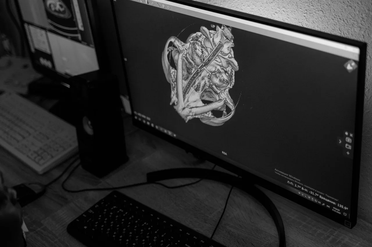
Some MRI scans use a contrast agent, usually based on gadolinium, injected into a vein to make certain tissues and blood vessels stand out. RadiologyInfo.org notes that gadolinium can be used safely in some patients who are allergic to the iodine-based contrast used in CT. Side effects are usually mild, but the NHS lists possible effects such as nausea, a skin rash, headache, a metallic taste or dizziness, and serious allergic reactions are rare. Tell staff about kidney problems, allergies or pregnancy before any contrast is given.
How to prepare
Preparation depends on the body part and whether contrast is planned. General steps include:
- Remove all metal: jewelry, watches, coins, keys, hairpins and clothing with metal fasteners. You may be given a gown.
- Complete a safety questionnaire about implants, devices, surgeries and any history of metal fragments in the eyes.
- Follow any fasting instructions if you are having abdominal or contrast imaging.
- Mention claustrophobia in advance so the team can plan support or discuss an open scanner.
Tip: speak up about metal and devices. Pacemakers, defibrillators, cochlear implants, some aneurysm clips and certain other devices can be unsafe in the magnetic field. MedlinePlus warns that the magnets can cause a piece of metal inside the body to move. When in doubt, tell the team before you enter the scan room. They would far rather check than assume.
What happens during the scan
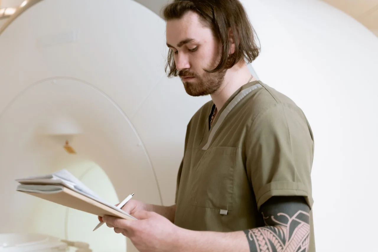
You lie on a motorized bed that slides into the tunnel of the scanner. A radiographer operates the machine from an adjoining room and stays in contact with you through an intercom. The NHS explains that the scanner produces loud bangs and clicks, so you are given ear protection such as headphones or earplugs. Staying still is important, because movement blurs the images. RadiologyInfo.org and MedlinePlus both note that a body MRI generally takes around 30 to 60 minutes, though some scans are shorter and others longer.
Safety and who should take extra care
The great advantage of MRI is that it uses no ionizing radiation. The trade-off is the powerful magnet, which is always on. That makes metal the main hazard rather than radiation. People with certain implanted electronic or metallic devices may not be able to have an MRI, or may need a specialized protocol. The NHS advises that patients disclose surgical implants, pacemakers or metal fragments before the scan.
Pregnancy is handled cautiously. MRI is often considered acceptable after the first trimester when needed, but gadolinium contrast is generally avoided in pregnancy unless truly necessary. Always share a possible pregnancy with your care team so they can weigh the options with you.
Important: this is general information, not medical advice. This article explains how MRI imaging generally works. It cannot tell you what your particular scan shows or what you should do next. Only your referring clinician or a radiologist, who can review your images alongside your full medical history, can interpret your results. Bring your questions to that appointment.
Understanding your results
A radiologist, a doctor specialized in reading images, examines your scan and writes a report for the clinician who ordered it. The NHS notes that MRI results typically take one to two weeks because the images need careful review. Reports often contain technical terms and measurements that sound alarming out of context. A described finding is not automatically a serious problem, and many incidental findings turn out to be harmless.
Your own doctor puts the report together with your symptoms, examination and other tests to decide what it means for you and whether any treatment or follow-up is needed. If you want to prepare for the financial side, our guide to MRI prices explains how these scans are typically billed.
Frequently asked questions
Does an MRI use radiation?
No. MRI uses a strong magnetic field and radio waves, not ionizing radiation. RadiologyInfo.org confirms that MRI does not use X-rays. This is one reason it is often chosen for repeated imaging or for younger patients when it can answer the clinical question.
How long does an MRI scan take?
Most scans take between about 15 and 60 minutes, depending on the body part and how many sequences are needed. A body MRI is commonly in the 30 to 60 minute range. Contrast studies can add time. Your clinic can give an estimate for your specific exam.
Is an MRI safe if I have metal in my body?
It depends on the type and location of the metal. Some implants and devices are MRI-compatible and some are not, and the strong magnet can move certain metal objects. Always disclose implants, pacemakers, aneurysm clips and any metal fragments so the team can confirm it is safe for you.
Will I be fully enclosed, and what if I am claustrophobic?
A standard MRI is a tunnel-shaped machine, and some scans require you to be well inside it. If you are claustrophobic, tell the clinic in advance. Options may include relaxation techniques, support from staff, medication in some cases, or an open MRI scanner where suitable.
Why is the machine so loud?
The knocking and buzzing sounds come from the scanner's gradient coils switching on and off rapidly during imaging. It is completely normal. You will be given ear protection such as headphones or earplugs, and you can usually listen to music or stay in contact with staff.
When will I get my results?
The images go to a radiologist for detailed review, and a report is sent to the doctor who ordered the scan. The NHS notes this often takes one to two weeks. Your own clinician will explain what the findings mean in the context of your health.
Sources: RadiologyInfo.org, Body MRI; NHS, MRI scan; MedlinePlus, Abdominal MRI.
More guides on MRI, CT & X-Ray
Have a question or a topic to suggest?
We read every message. Tell us what you would like to see next.
You may also like

MRI Scan: Uses, Procedure & Results
A plain-language guide to MRI scans: what they show, why a doctor might order one, how to prepare, what happens during the scan, and how results are read.
MRI of Pelvis: Uses, Procedure & Results
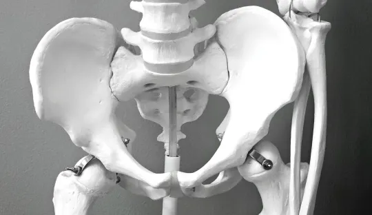
A pelvic MRI gives a detailed, radiation-free view of the bladder, reproductive organs, rectum and hip bones. Learn what it shows, how to prepare, contrast, and how results are interpreted.

MRI Prices 2026: With & Without Insurance
What an MRI really costs in 2026, with and without insurance. Understand the price ranges, what drives the cost, contrast fees, and practical ways to lower the bill.