Bone Density Scan: Uses and Results
Why DEXA is ordered, how the low-radiation scan works, and what T-scores and Z-scores mean, in plain language.
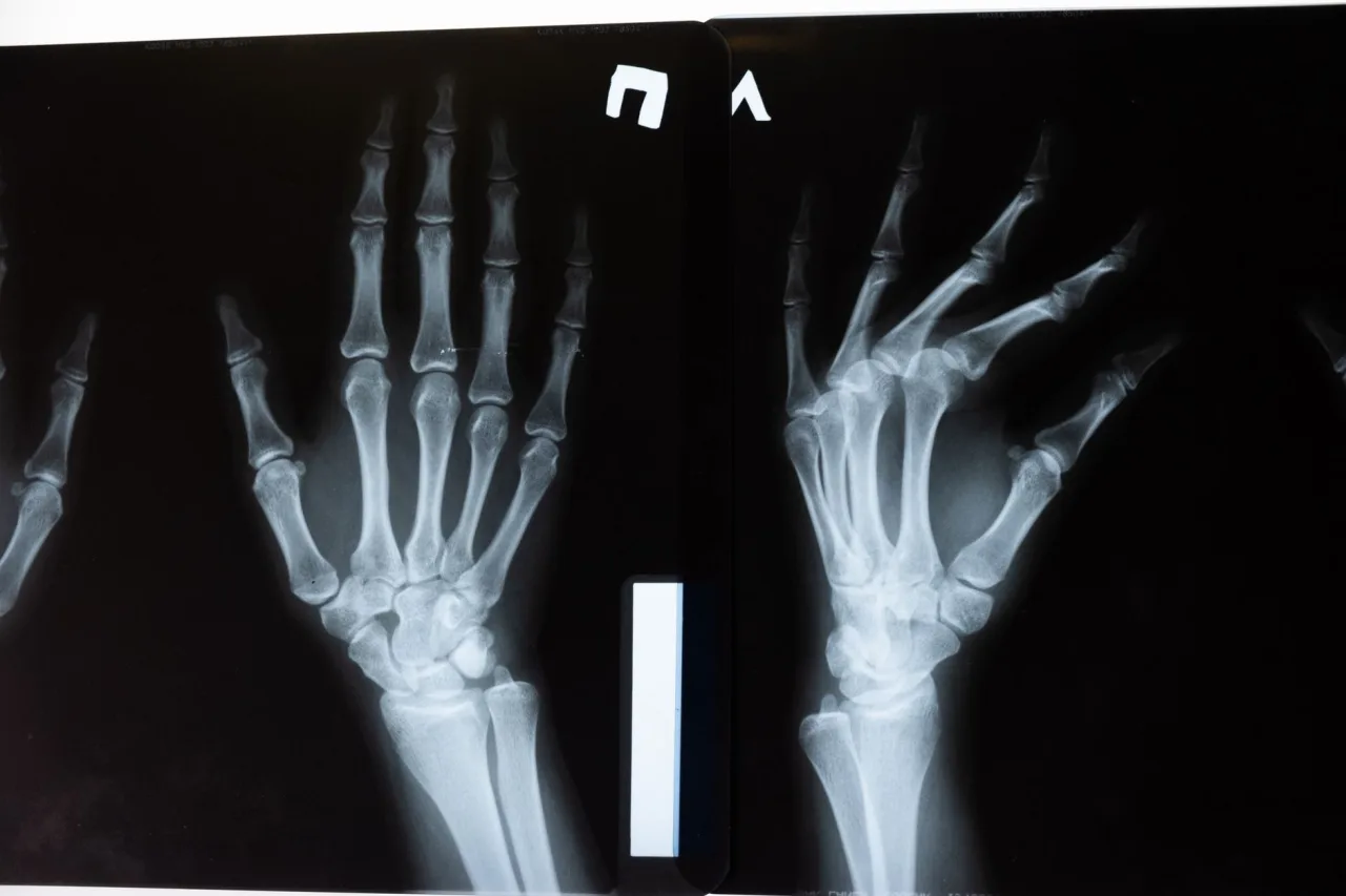
A bone density scan is a quick, low-radiation test that measures how strong your bones are and helps your doctor estimate your risk of a future fracture. It is the main tool used to diagnose osteoporosis and to track whether bone-strengthening treatment is working. This guide explains what the scan is, why it is ordered, how the procedure works, and how to read the numbers, in plain language.
The short version
- A bone density scan (usually a central DEXA) uses low-dose X-rays to measure bone mineral density, most often at the hip and lower spine.
- The radiation dose is very small, less than one-tenth of a standard chest X-ray and less than a day of natural background radiation.
- Results are reported as a T-score and a Z-score: a T-score of -2.5 or below indicates osteoporosis, and -1.1 to -2.4 indicates low bone mass (osteopenia).
- Only your own doctor can interpret your result in the context of your health, medicines, and fracture risk, so use this article to prepare good questions, not to self-diagnose.
What a bone density scan is
A bone density scan measures how much calcium and other minerals are packed into a section of bone. The denser the bone, the stronger it usually is, and the lower the chance it will break under everyday stress. The most common and most accurate version is central dual-energy X-ray absorptiometry, almost always shortened to central DEXA (sometimes written DXA).
According to MedlinePlus, a bone mineral density test measures how much calcium and other types of minerals are in an area of your bone, and the DEXA scan is the most common way to do it. RadiologyInfo.org describes DEXA as an enhanced form of X-ray technology used to measure bone loss, and calls it today's established standard for measuring bone mineral density.
There are two broad families of DEXA machine. Central DEXA scans your lower spine and hip while you lie on a padded table, and it is considered the best test for predicting fracture risk, especially of the hip. Peripheral DEXA uses smaller, sometimes portable machines to check bones in the wrist, finger, lower leg, or heel. Peripheral tests are useful for screening but are not as complete as a central scan.
Why a doctor orders one
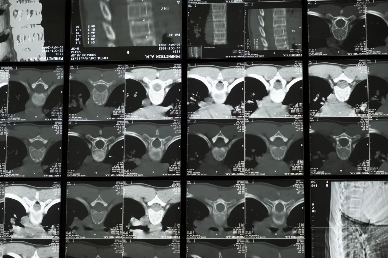
Bone density scans are most often used to diagnose or assess the risk of osteoporosis, a condition where bones become weak and more likely to fracture. Because bone loss usually causes no symptoms until a break happens, the scan is a way to find a problem early, while there is still time to act.
Reasons a clinician may recommend a scan include:
- Being a woman aged 65 or older, or a younger post-menopausal woman with risk factors.
- A previous low-impact fracture, for example breaking a bone after a minor fall.
- Long-term use of medicines that can thin bone, such as oral steroids.
- Conditions linked to bone loss, and lifestyle factors such as smoking, heavy alcohol use, or low body weight.
- Checking whether an osteoporosis treatment is working, by comparing scans over time.
The NHS notes that scans are often recommended for people over 50 with risk factors, and for some people under 50 who have risks such as smoking or a previous fracture. Your own history decides whether and when you need one, which is a conversation to have with your doctor.
How the procedure works
A DEXA scan is quick and painless. You stay fully clothed in most cases, as long as your clothing has no metal zips, buttons, or clasps over the area being scanned. You lie on your back on a flat, open table, so the test is a good option for people who feel uneasy in enclosed spaces.
The machine passes a thin, invisible beam of low-dose X-rays with two different energy levels through the bone. Special detectors measure how much of each beam passes through you. Because soft tissue and bone absorb the two energy levels differently, the software can subtract the soft tissue and calculate the bone mineral density on its own. For a hip and spine scan, you simply hold still while an arm above the table moves slowly over you. The whole appointment is usually over in about 10 to 30 minutes.
Preparing for your scan
- Stop taking calcium supplements for at least 24 hours before the scan, if your clinic advises it, because leftover supplement can affect the reading.
- Wear loose, comfortable clothing and leave metal jewellery at home.
- Tell the team if you may be pregnant. DEXA is generally avoided in pregnancy because any X-ray can affect an unborn baby.
- Mention any recent scan that used contrast dye or a radioactive tracer, as you may need to wait before a DEXA.
Radiation and safety
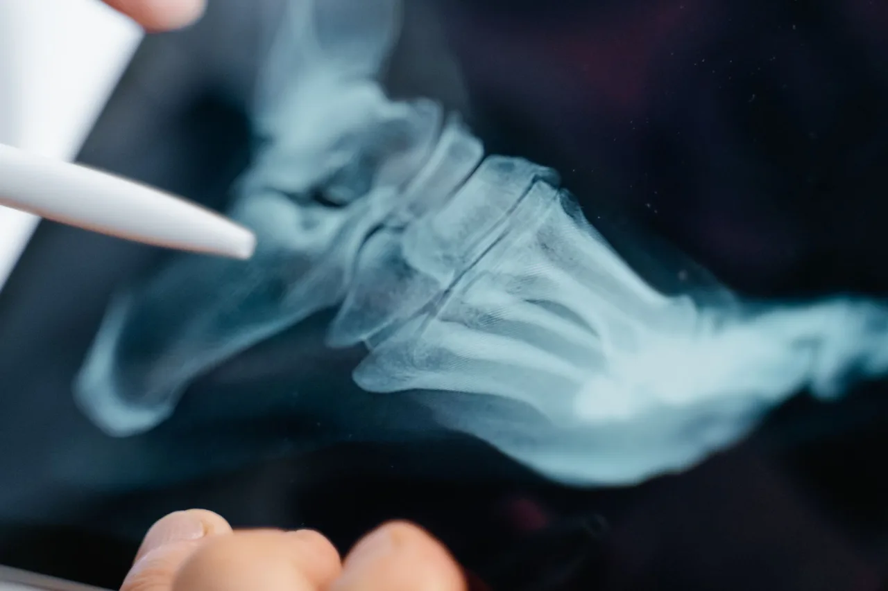
The radiation involved is very small. RadiologyInfo.org states the dose from a bone density scan is less than one-tenth the dose of a standard chest X-ray, and less than a day's exposure to natural background radiation. The NHS similarly explains that DEXA uses a much lower level of radiation than standard X-rays, which is why the test is considered very safe for most adults.
The main safety exception is pregnancy. Because X-rays can harm a developing baby, DEXA is not recommended for pregnant women, and staff will look for another approach if there is any chance you are pregnant. As with any radiology test, the goal is to use the smallest dose needed to get a useful answer, so always share your full history with the technologist.
Understanding your results
Your bone density is compared against reference groups and reported as two numbers.
- T-score. This compares your bone density with that of a healthy young adult at peak bone mass. Using the widely cited ranges from RadiologyInfo.org: a T-score of -1 and above is normal, -1.1 to -2.4 indicates low bone mass (osteopenia), and -2.5 or below indicates osteoporosis.
- Z-score. This compares your bone density with other people of your own age, size, and sex. A Z-score that is unusually low can prompt a doctor to look for a specific cause of bone loss beyond normal aging.
A more negative number means lower bone density and a higher estimated fracture risk. However, a score is only one part of the picture. Your doctor will combine it with your age, personal and family history, past fractures, and sometimes a separate fracture-risk calculator before deciding what, if anything, to do. That is why the same T-score can lead to different advice for two different people.
This is general information, not medical advice
This article explains bone density scans in general terms. It cannot tell you what your own results mean or what treatment you need. Always discuss your report, symptoms, and options with your doctor or the clinician who referred you, and follow their guidance.
How it compares with other imaging
A bone density scan answers a specific question about bone strength, and it is not the same as a general X-ray or a scan for another problem. A plain X-ray can show a fracture that has already happened but is not sensitive enough to measure gradual bone loss. A CT scan gives detailed cross-sections and can assess bone in specialised cases, but at a higher radiation dose than DEXA. If you want the bigger picture of how different tests fit together, our overview of radiology and what results mean is a helpful companion read.
Bone health also connects to other screening tests you may be offered around the same stage of life. A mammogram checks breast tissue rather than bone, but both are common preventive tests for adults and are sometimes booked together for convenience.
Finding a clinic and what to ask
Bone density scans are offered at hospital radiology departments, dedicated imaging centres, and some specialist clinics. When choosing where to go, look for accredited facilities with qualified technologists, and ask whether they can repeat your scan on the same machine in future so results stay comparable over time. Our guide to finding and comparing DEXA clinics walks through how to shortlist a good provider, and our DEXA scan explainer covers the technical side in more depth.
On cost, prices vary widely by country, provider, and whether you use insurance or pay privately, so always confirm the fee and any coverage with the clinic and your insurer before booking. Never assume a scan is covered without checking, and ask whether a referral is required.
Frequently asked questions
Does a bone density scan hurt?
No. A DEXA scan is painless and non-invasive. You lie still on an open table while a scanning arm passes over you, and nothing touches or enters your body. Most appointments take about 10 to 30 minutes.
How much radiation is in a DEXA scan?
Very little. RadiologyInfo.org states the dose is less than one-tenth that of a standard chest X-ray and less than a day of natural background radiation, which is why the test is considered very safe for most adults.
What T-score means I have osteoporosis?
Using the commonly cited ranges, a T-score of -1 and above is normal, -1.1 to -2.4 indicates low bone mass (osteopenia), and -2.5 or below indicates osteoporosis. Your doctor interprets this alongside your age and other risk factors before advising you.
Do I need to prepare for the scan?
Preparation is minimal. You may be asked to stop calcium supplements for at least 24 hours beforehand, to wear clothing without metal, and to avoid the test if you may be pregnant. Follow the specific instructions your clinic gives you.
How often should the scan be repeated?
That depends on your first result and your risk factors, and it is decided by your doctor. Some people are rescanned after a couple of years to monitor change or check that treatment is working, ideally on the same machine so the numbers can be compared fairly.
Can I have a DEXA scan if I am pregnant?
Generally no. Because X-rays can harm an unborn baby, DEXA is not recommended during pregnancy. Tell the team if there is any chance you are pregnant so they can consider alternatives or reschedule.
More guides on Mammogram & Bone Density
Have a question or a topic to suggest?
We read every message. Tell us what you would like to see next.
You may also like
DEXA Scan: Uses, Procedure and Results
A DEXA scan is the standard test for measuring bone density and checking for osteoporosis. Here is why it is ordered, how the scan works, and what your T-score means, with a reminder to always discuss results with your doctor.
DEXA Scan Near Me: Choose a Clinic
A DEXA scan is one test where the clinic you pick has a lasting effect, because results are compared over years. Learn how to choose a good bone density center.

Mammogram: Uses, Procedure & Results
Understand the mammogram: screening versus diagnostic, how to prepare, what the exam feels like, radiation safety, and how your results are handled.