DEXA Scan: Uses, Procedure and Results
Why a bone density scan is ordered, how it works, the small radiation dose, and how to read a T-score, in plain language.
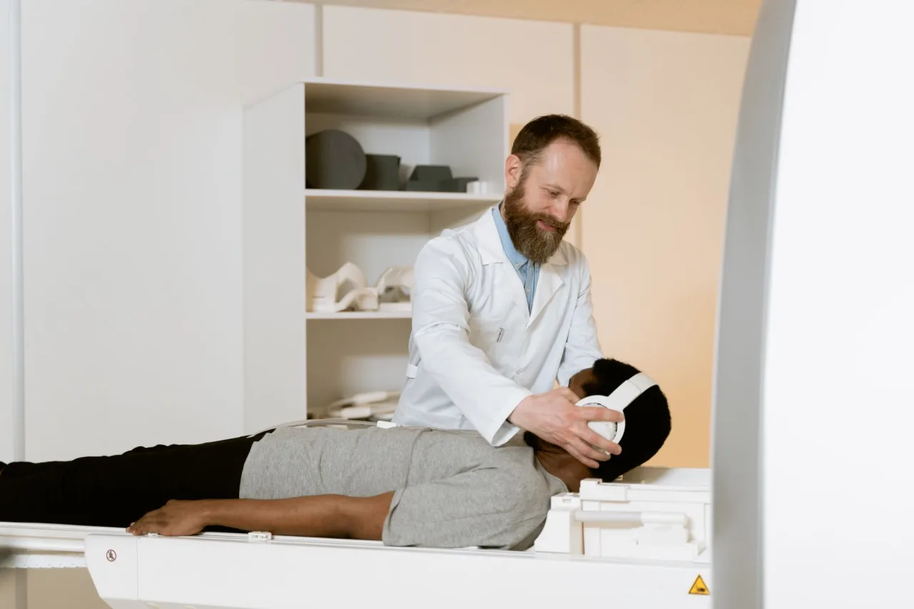
A DEXA scan (also written DXA, and short for dual-energy x-ray absorptiometry) is the standard test doctors use to measure bone density and check for osteoporosis. It is quick, painless, and uses a very small amount of radiation. This guide explains why a DEXA scan is ordered, how it works, what the T-score on your report means, and why the numbers should always be read alongside your own doctor.
The short version
- A DEXA scan measures how strong and dense your bones are, most often at the hip and lower spine.
- The radiation dose is tiny, less than one-tenth of a standard chest x-ray and less than a normal day of natural background radiation.
- Results are given as a T-score: about -1 and above is normal, -1.1 to -2.4 is low bone mass (osteopenia), and -2.5 or below suggests osteoporosis.
- Your T-score is only part of the picture. A doctor weighs it with your age, history, and fracture risk before deciding anything.
What a DEXA scan is
DEXA stands for dual-energy x-ray absorptiometry. The scanner sends two low-dose x-ray beams of different energy levels through the bone. Because bone and soft tissue absorb the two beams differently, the machine can calculate how much mineral is packed into a given area of bone. That measurement is called bone mineral density, and it is the best-established way to estimate bone strength and the risk of fracture.
The test usually focuses on the lower spine and the hips, because these are the sites where fractures cause the most harm and where the reading predicts risk most reliably. Some clinics also scan the forearm. If you want the wider context of how imaging tests fit together, our overview of radiology as a whole is a helpful companion read.
Why a doctor orders one
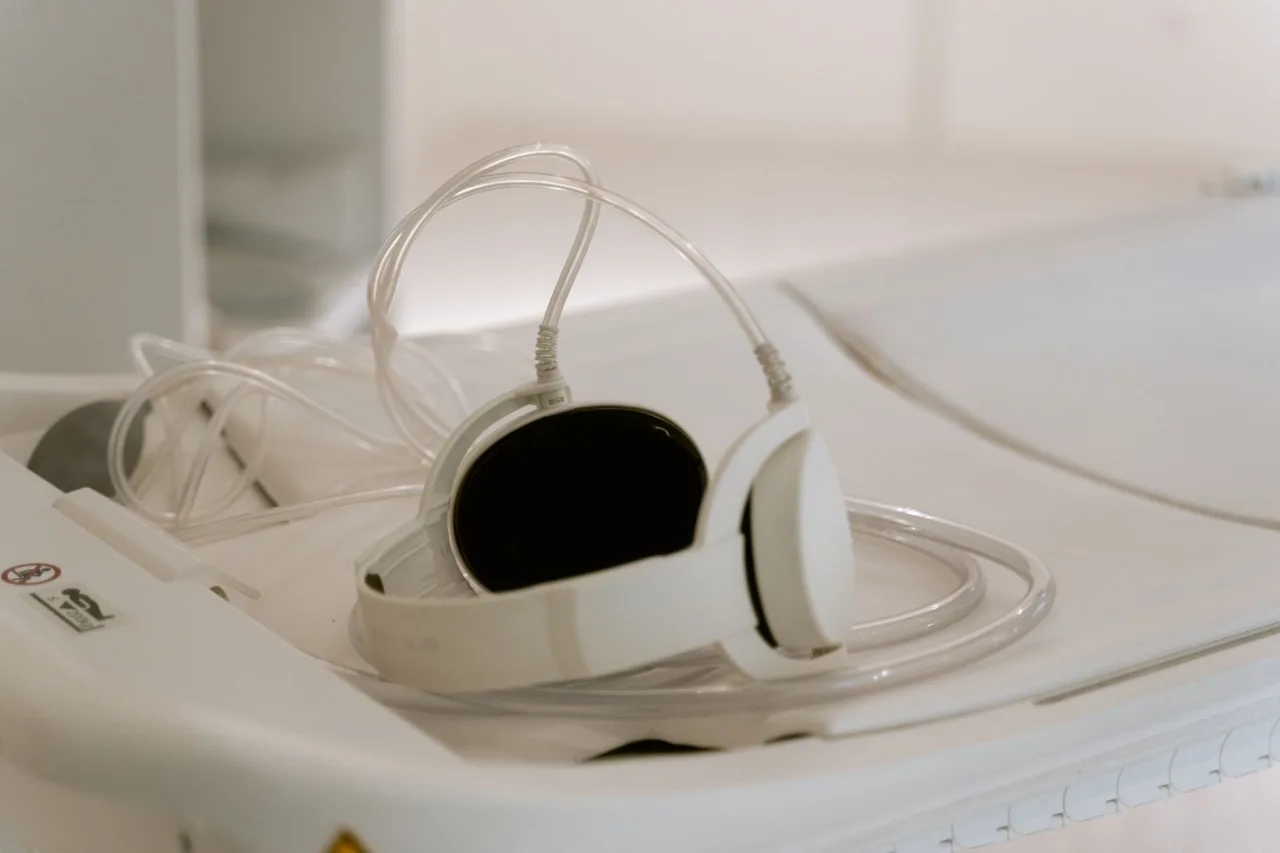
Doctors order a DEXA scan to diagnose osteoporosis before a bone breaks, to estimate your fracture risk, and to monitor whether treatment is working. According to RadiologyInfo.org, common reasons include:
- Being a woman past menopause who is not taking oestrogen.
- Being older, since bone density falls with age in both men and women.
- A personal history of fractures, or a parent who broke a hip.
- Long-term use of medicines that thin bone, such as corticosteroids.
- Conditions linked to bone loss, including an overactive thyroid or parathyroid.
The point of the scan is prevention. Osteoporosis has no symptoms until a bone breaks, so measuring density early gives you and your doctor a chance to act before that happens.
How the scan works, step by step
A DEXA scan is one of the more comfortable imaging tests. You stay dressed as long as your clothing has no metal zips, buttons, or clasps in the scan area.
- You lie on your back on a padded table. A large arm passes above you without touching you.
- For a hip and spine scan, a positioner may be placed under your legs to flatten the lower spine and rotate the hip.
- You stay still and, for very short moments, hold your breath so the image is sharp.
- The scanning arm moves slowly over the area while the detector records the readings.
The whole appointment usually takes 10 to 30 minutes depending on the equipment and how many sites are measured. There is no injection, no tunnel, and no dye.
How to prepare
- Eat and drink normally. No fasting is needed.
- Stop calcium supplements for at least 24 hours before the scan, because undissolved tablets can distort the reading.
- Wear loose, comfortable clothing without metal fastenings.
- Tell the clinic if you might be pregnant, or if you have had a recent barium study or contrast injection, as these can affect results.
Radiation and safety
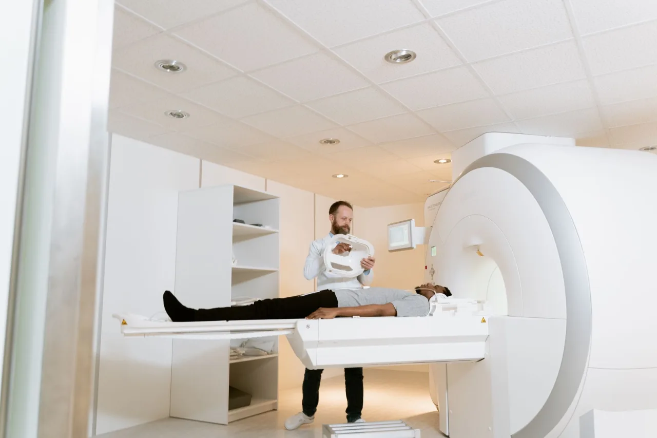
The radiation from a DEXA scan is very low. RadiologyInfo.org notes the dose is less than one-tenth of a standard chest x-ray and less than a day of the natural background radiation everyone is exposed to. The NHS makes the same point, adding that the dose is low enough that the radiographer can stay in the room during the scan.
The main safety exception is pregnancy. Because even small doses of radiation are avoided during pregnancy where possible, tell your clinic before the scan if there is any chance you are pregnant, so they can decide whether to postpone.
Understanding your T-score and Z-score
Your DEXA report gives two main numbers. The T-score compares your bone density to that of a healthy young adult. The Z-score compares it to other people of your own age and sex. For diagnosing osteoporosis after menopause and in older men, the T-score is the one that matters most.
- -1.0 and above: normal bone density.
- -1.1 to -2.4: low bone mass, sometimes called osteopenia.
- -2.5 and below: a result consistent with osteoporosis.
These thresholds come from World Health Organization criteria and are quoted by RadiologyInfo.org. A lower, more negative number means thinner bone. But a score never stands alone. Two people with the same T-score can have very different fracture risks once age, weight, smoking, and past fractures are taken into account, which is why tools like FRAX combine the density reading with those other factors.
It also helps to know what a DEXA scan does not measure. It reports how much mineral is packed into a flat, two-dimensional area of bone, which is an excellent guide to strength but not a complete one. It does not directly capture bone quality, the internal architecture that also affects how easily a bone breaks. That is another reason your clinician reads the number alongside the bigger picture rather than treating it as a verdict on its own. In younger adults and children, the Z-score is used instead of the T-score, because comparing a young person to a healthy young adult would not be meaningful.
Some clinics also offer peripheral DEXA, a smaller machine that measures the wrist, finger, or heel. These devices are portable and useful for screening, but a central scan of the hip and spine remains the reference standard for diagnosis and for tracking change over time.
This is general information, not medical advice. A DEXA result can only be interpreted properly by a clinician who knows your full history. Do not start, stop, or change any treatment based on a number you read here. Always discuss your report with your doctor or the specialist who ordered the scan.
What happens after the scan
A radiologist, a doctor trained to interpret imaging, reviews the scan and sends a report to the doctor who requested it. That doctor then talks the result through with you. Depending on the findings, the conversation might cover diet and vitamin D, weight-bearing exercise, reducing fall risk at home, or medicines that slow bone loss.
If you are being monitored, follow-up scans are usually spaced a year or two apart, because bone density changes slowly and scanning too often does not add useful information. Comparing scans is only reliable when they are done on the same type of machine, so try to return to the same clinic where possible.
DEXA compared with other imaging
People sometimes confuse a DEXA scan with other tests. It is not the same as a mammogram, which images breast tissue, nor a CT scan, which uses far more radiation to build detailed cross-sections. DEXA is a low-dose, targeted measurement of bone. The professionals who perform these tests train in different specialties, and if the imaging career path interests you, our mammographer career guide covers a related role.
When you are ready to book, comparing accredited providers matters more than picking the nearest name. Our guide to choosing a diagnostic centre near you explains what to look for.
Frequently asked questions
Does a DEXA scan hurt?
No. A DEXA scan is painless. You simply lie still on a table while a scanning arm passes over you. There are no injections, no enclosed tunnel, and nothing touches the area being scanned.
How much radiation does a DEXA scan use?
Very little. The dose is less than one-tenth of a standard chest x-ray and less than the natural background radiation you receive in a single day, according to RadiologyInfo.org. The dose is low enough that staff can remain in the room.
What does a T-score of -2.5 mean?
A T-score of -2.5 or lower is the threshold that is consistent with osteoporosis. However, only your doctor can interpret what it means for you, because fracture risk also depends on age, previous fractures, and other factors. Discuss the result with the clinician who ordered the scan.
How often should a DEXA scan be repeated?
When used to monitor bone, follow-up scans are usually one to two years apart, since bone density changes slowly. Your doctor decides the interval based on your risk and any treatment you are taking. For reliable comparison, repeat scans should be done on the same machine.
Do I need to prepare for a DEXA scan?
Preparation is minimal. Eat and drink normally, but avoid calcium supplements for at least 24 hours beforehand, and wear clothing without metal fastenings. Tell the clinic if you might be pregnant or have recently had a contrast injection or barium study.
More guides on Mammogram & Bone Density
Have a question or a topic to suggest?
We read every message. Tell us what you would like to see next.
You may also like

Mammogram: Uses, Procedure & Results
Understand the mammogram: screening versus diagnostic, how to prepare, what the exam feels like, radiation safety, and how your results are handled.
CT Scan: Uses, Procedure and Results
A CT scan, or CAT scan, uses x-rays and a computer to create detailed cross-sectional images of the body. Here is why doctors order one, how the procedure works, what contrast dye does, and how the radiation compares.

Mammographer: 2026 Career Guide
A 2026 career guide to becoming a mammographer: education, ARRT certification, work settings, salary ranges, and the job outlook, with cited sources.